コンテンツへスキップ
Merck

MABC32

Sigma-Aldrich

抗p62(セクエストソーム-1)抗体、クローン11C9.2

clone 11C9.2, from mouse

別名:

sequestosome 1, EBI3-associated protein of 60 kDa, Paget disease of bone 3, phosphotyrosine independent ligand for the Lck SH2 domain p62, oxidative stress induced like, EBI3-associated protein p60, Phosphotyrosine-independent ligand for the Lck SH2 doma

ログイン組織・契約価格を表示する


About This Item

UNSPSCコード:
12352203
eCl@ss:
32160702
NACRES:
NA.41

由来生物

mouse

品質水準

抗体製品の状態

purified antibody

抗体製品タイプ

primary antibodies

クローン

11C9.2, monoclonal

化学種の反応性

mouse, rat, human

テクニック

flow cytometry: suitable
immunocytochemistry: suitable
western blot: suitable

アイソタイプ

IgMκ

NCBIアクセッション番号

UniProtアクセッション番号

輸送温度

wet ice

ターゲットの翻訳後修飾

unmodified

遺伝子情報

human ... SQSTM1(8878)

関連するカテゴリー

詳細

p62は、別名セクエストソーム-1であり、62 kDaのras-GAP関連タンパク質です。p62はsrcファミリーチロシンキナーゼに結合すると考えられており、活性化srcファミリーキナーゼとその下流エフェクターとのリンカー作用も行う可能性があります。p62にはRNA結合領域があるため、RNA結合能を有する可能性があります。p62は、転写因子NF-κBの活性化と関連しており、細胞分化、免疫応答、およびカリウムチャネル調節に関与しています。p62は最近、外因性アポトーシスと関連付けられており、腫瘍形成において重要な役割を果たしている可能性があります。

免疫原

ヒトp62(セクエストソーム-1)に相当するGSTタグ付きリコンビナントタンパク質。

アプリケーション

この抗p62(セクエストソーム-1)抗体、クローン11C9.2を用いたp62(セクエストソーム-1)の検出は、WB、FC、ICでの使用が検証されています。
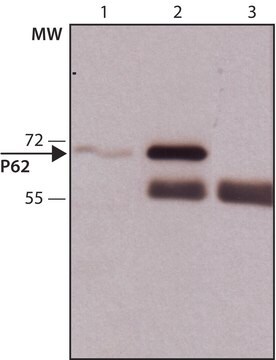
ウェスタンブロッティング:0.001 µg/mLで使用、10 µgのPC12細胞ライセート中のp62(セクエストソーム-1)を検出できます。

フローサイトメトリー:1.0 µgで使用、固定し透過処理したHeLa細胞の染色において p62(セクエストソーム-1)を検出できます。

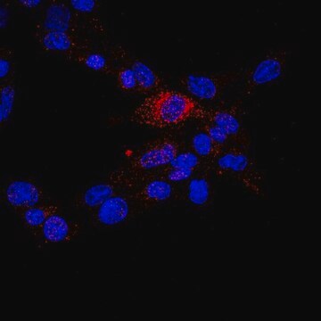
免疫細胞染色:希釈倍率1:500で使用、NIH/3T3、A431、HeLa細胞のp62 (セクエストソーム-1)を検出できます。
研究カテゴリー
アポトーシス・癌
研究サブカテゴリー
アポトーシス-追加

品質

A431細胞ライセートのウェスタンブロットで評価されています。

ウェスタンブロッティング:0.001 µg/mLで使用、10 µgのA431細胞ライセート中のp62(セクエストソーム-1)を検出できます。

ターゲットの説明

約47-60 kDa、実測。
このタンパク質の算出した分子量は47 kDaであり、修飾による38 kDaのアイソフォームもあります。このタンパク質は一部のライセートでは最高約60 kDaのものが認められることがあります。

物理的形状

PBSおよび0.05%アジ化ナトリウムを含むバッファー中の精製マウスモノクローナルIgMκ。
フォーマット:精製

保管および安定性

2~8°Cで受領日から1年間安定です。

アナリシスノート

コントロール
A431細胞ライセート

免責事項

メルクのカタログまたは製品に添付されたメルクのその他の文書に記載されていない場合、メルクの製品は研究用途のみを目的としているため、他のいかなる目的にも使用することはできません。このような目的としては、未承認の商業用途、in vitroの診断用途、ex vivoあるいはin vivoの治療用途、またはヒトあるいは動物へのあらゆる種類の消費あるいは適用などがありますが、これらに限定されません。

適切な製品が見つかりませんか。  

製品選択ツール.をお試しください

保管分類コード

12 - Non Combustible Liquids

WGK

WGK 2

引火点(°F)

Not applicable

引火点(℃)

Not applicable


試験成績書(COA)

製品のロット番号・バッチ番号を入力して、試験成績書(COA) を検索できます。ロット番号・バッチ番号は、製品ラベルに「Lot」または「Batch」に続いて記載されています。

以前この製品を購入いただいたことがある場合

文書ライブラリで、最近購入した製品の文書を検索できます。

文書ライブラリにアクセスする

AMPK involvement in endoplasmic reticulum stress and autophagy modulation after fatty liver graft preservation: a role for melatonin and trimetazidine cocktail.
Zaouali, Mohamed Amine, et al.
Journal of Pineal Research (2013)
Huimin Yan et al.
Journal of rheumatic diseases and treatment, 1(1), 5-5 (2015-06-30)
Autophagy plays a central role in various disease processes. However, its contribution to inflammatory arthritides such as rheumatoid arthritis (RA) is unclear. We observed that autophagy is engaged in the K/BxN serum transfer model of RA but autophagic flux is
Michalis Papadakis et al.
Nature medicine, 19(3), 351-357 (2013-02-26)
Previous attempts to identify neuroprotective targets by studying the ischemic cascade and devising ways to suppress it have failed to translate to efficacious therapies for acute ischemic stroke. We hypothesized that studying the molecular determinants of endogenous neuroprotection in two
Mallika Valapala et al.
Autophagy, 10(3), 480-496 (2014-01-29)
In phagocytic cells, including the retinal pigment epithelium (RPE), acidic compartments of the endolysosomal system are regulators of both phagocytosis and autophagy, thereby helping to maintain cellular homeostasis. The acidification of the endolysosomal system is modulated by a proton pump
Elisa Teyssou et al.
Neurobiology of aging, 58, 239-239 (2017-07-19)
Mutations in UBQLN2 have been associated with rare cases of X-linked juvenile and adult forms of amyotrophic lateral sclerosis (ALS) and ALS linked to frontotemporal dementia (FTD). Here, we report 1 known (c.1489C>T, p.Pro497Ser, P497S) and 3 novel (c.1481C>T, p.Pro494Leu

資料

Autophagy is a highly regulated process that is involved in cell growth, development, and death. In autophagy cells destroy their own cytoplasmic components in a very systematic manner and recycle them.

Autophagy is a highly regulated process that is involved in cell growth, development, and death. In autophagy cells destroy their own cytoplasmic components in a very systematic manner and recycle them.

ライフサイエンス、有機合成、材料科学、クロマトグラフィー、分析など、あらゆる分野の研究に経験のあるメンバーがおります。.

製品に関するお問い合わせはこちら(テクニカルサービス)