
284M-9
Microphthalmia Transcription Factor (MiTF) (C5/D5) Mouse Monoclonal Antibody
About This Item
Prodotti consigliati


Origine biologica
mouse
Livello qualitativo
100
500
Coniugato
unconjugated
Forma dell’anticorpo
culture supernatant
Tipo di anticorpo
primary antibodies
Clone
C5/D5, monoclonal
Descrizione
For In Vitro Diagnostic Use in Select Regions (See Chart)
Stato
buffered aqueous solution
Reattività contro le specie
human
Confezionamento
vial of 0.1 mL concentrate (284M-94)
vial of 0.5 mL concentrate (284M-95)
bottle of 1.0 mL predilute (284M-97)
vial of 1.0 mL concentrate (284M-96)
bottle of 7.0 mL predilute (284M-98)
Produttore/marchio commerciale
Cell Marque®
tecniche
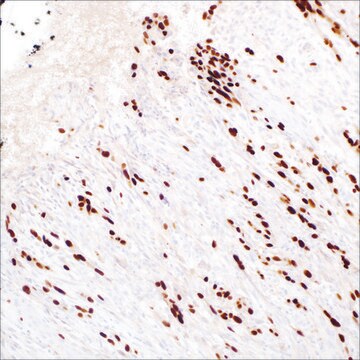
immunohistochemistry (formalin-fixed, paraffin-embedded sections): 1:100-1:500
Isotipo
IgG1
Controllo
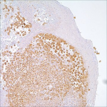
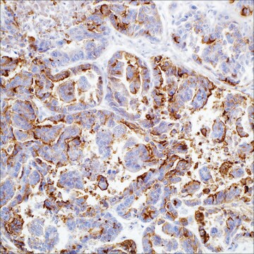
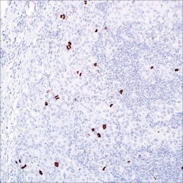
melanoma
Condizioni di spedizione
wet ice
Temperatura di conservazione
2-8°C
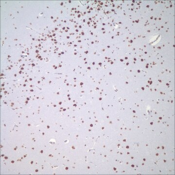
Visualizzazione
nuclear
Informazioni sul gene
human ... MITF(4286)
Categorie correlate
Descrizione generale
Qualità
 IVD |  IVD |  IVD |  RUO |
Linkage
Stato fisico
Nota sulla preparazione
Altre note
Note legali
Non trovi il prodotto giusto?
Prova il nostro Motore di ricerca dei prodotti.
Scegli una delle versioni più recenti:
Certificati d'analisi (COA)
Non trovi la versione di tuo interesse?
Se hai bisogno di una versione specifica, puoi cercare il certificato tramite il numero di lotto.
Possiedi già questo prodotto?
I documenti relativi ai prodotti acquistati recentemente sono disponibili nell’Archivio dei documenti.
Articoli
Immunohistochemistry (IHC) techniques and applications have greatly improved, dermatopathology is still largely based on H&E stained slides.This paper outlines ways in which IHC antibodies can be utilized for dermatopathology.
Il team dei nostri ricercatori vanta grande esperienza in tutte le aree della ricerca quali Life Science, scienza dei materiali, sintesi chimica, cromatografia, discipline analitiche, ecc..
Contatta l'Assistenza Tecnica.