12209136001
Roche
TeloTAGGG™ Telomere Length Assay
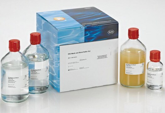
sufficient for ≤50 reactions, kit of 1 (15 components), suitable for cell culture
Sinonimo/i:
telomere
Autenticatiper visualizzare i prezzi riservati alla tua organizzazione & contrattuali
About This Item
Codice UNSPSC:
41105500
Prodotti consigliati
impiego
sufficient for ≤50 reactions
Livello qualitativo
Confezionamento
kit of 1 (15 components)
Produttore/marchio commerciale
Roche
tecniche
cell culture | mammalian: suitable
Temperatura di conservazione
−20°C
Categorie correlate
Descrizione generale
Telomeres, the specialized DNA protein structures located at the end of eukaryotic chromosomes, consist of small, tandemly repeated DNA sequences. Numerous telomere sequences have been identified that display very few sequence variations, even between phylogenetically divergent organisms such as Tetrahymena (sequence: TTGGGG) and humans (sequence: TTAGGG).
Because DNA polymerase is unable to replicate the very ends of linear DNA, it was suggested that chromosomal ends progressively shorten with each replication cycle (called the “end-replication problem”). This phenomenon, which has been demonstrated in vitro and in vivo, seems to be linked to the limited proliferative capacity of normal somatic cells (“mitotic clock”). Since germ-line cells, stem cells, and tumor cells all exhibit a prolonged or even infinite life span, it was suggested that these cells must possess a particular mechanism for maintaining telomere length.
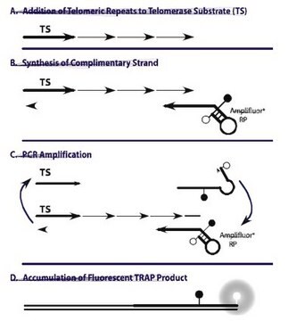
Maintaining stable telomere length is associated with the activation of telomerase. This enzyme is a ribonucleoprotein that compensates for the loss of telomeric DNA by adding repeat sequences to the chromosome ends, using its intrinsic RNA component as a template for DNA synthesis.
Telomeres play an essential role in the stable maintenance of eukaryotic chromosomes within a cell by specifically binding to structural proteins. These proteins cap the ends of linear chromosomes, preventing nucleolytic degradation, end-to-end fusion, irregular recombination, and other events that are normally lethal to a cell.
Analysis of telomere length in research samples of human peripheral blood mononuclear cells reveals that telomere length decreases with increased age in the donor, reflecting the replicative history of those cells. In several disorders (e.g., Down′s syndrome, ataxia telangiectasia, and HIV infection), accelerated telomere loss has been described, suggesting the reduction in telomere length may be related to the immune dysfunction associated with these disorders. This kit is intended to increase scientific knowledge about these relationships.
Assay time: Approximately 18 hours
Sample material: Cell cultures and other biological samples
Nonradioactive chemiluminescent assay to determine telomere length.
The kit utilizes Southern analysis of terminal restriction fragments (TRF) that are obtained by the digestion of genomic DNA using frequently cutting restriction enzymes.
Step 1: Digestion of genomic DNA
Purified genomic DNA is digested by an optimized mixture of frequently cutting restriction enzymes. The enzymes have been selected in such a way that telomeric DNA and sub-telomeric DNA is not cut. This is due to the special sequence characteristics of the repeats. Non-telomeric DNA is digested to low molecular-weight fragments.
Step 2: Gel electrophoresis and Southern blotting
Following DNA digestion, the DNA fragments are separated by gel electrophoresis, then transferred to a nylon membrane by Southern blotting.
Step 3: Hybridization and chemiluminescence detection
The blotted DNA fragments are hybridized to a digoxigenin (DIG)-labeled probe that is specific for telomeric repeats, then incubated with a DIG-specific antibody covalently coupled to alkaline phosphatase. Finally, the immobilized telomere probe is visualized by a highly sensitive chemiluminescent substrate for alkaline phosphatase, CDP-Star. The average TRF length can be determined by comparing the signals to a molecular-weight standard.
Applicazioni
The TeloTAGGG™ Telomere Length Assay is designed for use in the following life science research applications:
- Sensitive detection of telomeric DNA (telomeric sequence: TTAGGG) in cell cultures and other biological research samples
- Determination of the telomere length of DNA in cell cultures and other biological research samples
Caratteristiche e vantaggi
- Safe: Nonradioactive
- Flexible: Detects telomeres from a variety of organisms, including humans and mice
Confezionamento
1 kit containing 15 components.
Sequenza
Sequence and length of the probe are confidential. The DIG labeled MW in the Kit is a mixture of DIG MW III and VII
Nota sulla preparazione
Working concentration: Working concentration of conjugate depends on application and substrate
The following concentrations should be taken as a guideline:
Working solution: TAE buffer
0.04 M Tris-acetate, 0.001 M EDTA, pH 8.0
HCl solution
0.25 M HCl
For a 200 cm2 blot about 250 ml of solution are needed.
Denaturation solution
0.5 M NaOH, 1.5 M NaCl
For a 200 cm2 blot about 500 ml of solution are needed.
Neutralization solution
0.5 M Tris-HCl, 3 M NaCl, pH 7.5
For a 200 cm2 blot about 500 ml of solution are needed.
20x SSC
3 M NaCl, 0.3 M Sodium citrate, pH 7.0
2x SSC
Dilute 20x SSC (Solution 5) 1:10 with autoclaved, redistilled water.
DIG Easy Hyb granules
Reconstitute the granules (Bottle 8) with 64 ml autoclaved, redistilled water and incubate at 37 °C until complete reconstitution. Prepare the solution several hours before use.
Stringent wash buffer I
2x SSC, 0.1% SDS
Stringent wash buffer II
0.2x SSC, 0.1% SDS
Washing buffer, 1x
Dilute an appropriate volume of washing buffer, 10x (Bottle 10) 1:10 with autoclaved, redistilled water.
Blocking solution, 1x
Dilute an appropriate volume of blocking buffer, 10x (Bottle 12) 1:10 with maleic acid buffer, 1x (Solution 12).
Maleic acid buffer, 1×
Dilute an appropriate volume of maleic acid buffer, 10x (Bottle 11) 1:10 with autoclaved, redistilled water.
Anti-DIG-AP, working solution
For reducing background by aggregated antibody, please spin vial for 5 min at 13,000 rpm before use.
Dilute an appropriate volume of Anti-DIG-AP (Bottle 13) with blocking solution, 1x (Solution 11) to a final concentration of 75 mU/ml (1:10,000).
Detection buffer, 1x
Dilute an appropriate volume of detection buffer, 10x (Bottle 14) 1:10 with autoclaved, redistilled water.
Storage conditions (working solution): TAE buffer:
Stable at 15 to 25 °C
HCl solution
Stable at 15 to 25 °C
Denaturation solution
Stable at 15 to 25 °C
Neutralization solution
Stable at 15 to 25 °C
20x SSC
Stable at 15 to 25 °C
2x SSC
Stable at 15 to 25 °C
DIG Easy Hyb
Stable at 15 to 25 °C for 3 months
Stringent wash buffer I
Stable at 15 to 25 °C
Stringent wash buffer II
stable at 15 to 25 °C
Washing buffer, 1x
Stable at 15 to 25 °C
Blocking solution, 1x
Prepare just before use. Do not store
Maleic acid buffer, 1×
Stable at 15 to 25 °C
Anti-DIG-AP, working solution
Prepare just before use. Do not store
Detection buffer, 1x
Stable at 15 to 25 °C
The following concentrations should be taken as a guideline:
- Dot blot: 100 ng/ml
- ELISA: 100 ng/ml
- Western blot: 100 ng/ml
Working solution: TAE buffer
0.04 M Tris-acetate, 0.001 M EDTA, pH 8.0
HCl solution
0.25 M HCl
For a 200 cm2 blot about 250 ml of solution are needed.
Denaturation solution
0.5 M NaOH, 1.5 M NaCl
For a 200 cm2 blot about 500 ml of solution are needed.
Neutralization solution
0.5 M Tris-HCl, 3 M NaCl, pH 7.5
For a 200 cm2 blot about 500 ml of solution are needed.
20x SSC
3 M NaCl, 0.3 M Sodium citrate, pH 7.0
2x SSC
Dilute 20x SSC (Solution 5) 1:10 with autoclaved, redistilled water.
DIG Easy Hyb granules
Reconstitute the granules (Bottle 8) with 64 ml autoclaved, redistilled water and incubate at 37 °C until complete reconstitution. Prepare the solution several hours before use.
Stringent wash buffer I
2x SSC, 0.1% SDS
Stringent wash buffer II
0.2x SSC, 0.1% SDS
Washing buffer, 1x
Dilute an appropriate volume of washing buffer, 10x (Bottle 10) 1:10 with autoclaved, redistilled water.
Blocking solution, 1x
Dilute an appropriate volume of blocking buffer, 10x (Bottle 12) 1:10 with maleic acid buffer, 1x (Solution 12).
Maleic acid buffer, 1×
Dilute an appropriate volume of maleic acid buffer, 10x (Bottle 11) 1:10 with autoclaved, redistilled water.
Anti-DIG-AP, working solution
For reducing background by aggregated antibody, please spin vial for 5 min at 13,000 rpm before use.
Dilute an appropriate volume of Anti-DIG-AP (Bottle 13) with blocking solution, 1x (Solution 11) to a final concentration of 75 mU/ml (1:10,000).
Detection buffer, 1x
Dilute an appropriate volume of detection buffer, 10x (Bottle 14) 1:10 with autoclaved, redistilled water.
Storage conditions (working solution): TAE buffer:
Stable at 15 to 25 °C
HCl solution
Stable at 15 to 25 °C
Denaturation solution
Stable at 15 to 25 °C
Neutralization solution
Stable at 15 to 25 °C
20x SSC
Stable at 15 to 25 °C
2x SSC
Stable at 15 to 25 °C
DIG Easy Hyb
Stable at 15 to 25 °C for 3 months
Stringent wash buffer I
Stable at 15 to 25 °C
Stringent wash buffer II
stable at 15 to 25 °C
Washing buffer, 1x
Stable at 15 to 25 °C
Blocking solution, 1x
Prepare just before use. Do not store
Maleic acid buffer, 1×
Stable at 15 to 25 °C
Anti-DIG-AP, working solution
Prepare just before use. Do not store
Detection buffer, 1x
Stable at 15 to 25 °C
Altre note
For life science research only. Not for use in diagnostic procedures.
Note legali
Manufactured under license from Geron Corporation.
TeloTAGGG is a trademark of Roche
Solo come componenti del kit
N° Catalogo
Descrizione
- Hinf I (30μl) 40U/μl
- Rsa I (30μl) 40U/μl
- Digestion Buffer 10x concentrated
- Water, nuclease-free
- Control DNA 150μl
- DIG Molecular Weight Marker 40 μl
- Loading Buffer 5x concentrated
- DIG Easy Hyb Granules
- Telomerase Probe
- Washing Buffer 10x concentrated
- Maleic Acid Buffer 10x concentrated
- Blocking Buffer 10x concentrated
- Anti-DIG-AP antibody
- Detection Buffer 10x concentrated
- Substrate Solution (CDP-Star) ready-to-use
Vedi tutto (15)
Avvertenze
Warning
Indicazioni di pericolo
Consigli di prudenza
Classi di pericolo
Eye Irrit. 2 - Skin Irrit. 2
Codice della classe di stoccaggio
12 - Non Combustible Liquids
Classe di pericolosità dell'acqua (WGK)
WGK 3
Punto d’infiammabilità (°F)
does not flash
Punto d’infiammabilità (°C)
does not flash
Certificati d'analisi (COA)
Cerca il Certificati d'analisi (COA) digitando il numero di lotto/batch corrispondente. I numeri di lotto o di batch sono stampati sull'etichetta dei prodotti dopo la parola ‘Lotto’ o ‘Batch’.
Possiedi già questo prodotto?
I documenti relativi ai prodotti acquistati recentemente sono disponibili nell’Archivio dei documenti.
G Yang et al.
Oncogene, 26(10), 1492-1498 (2006-09-06)
The risk of developing ovarian cancer is about 1% over a lifetime, but it is the most deadly gynecologic cancer, in part due to lack of diagnostic markers for early-stage disease and cell model system for studying early neoplastic changes.
Matthew Parker et al.
Genome biology, 13(12), R113-R113 (2012-12-13)
Telomeres are the protective arrays of tandem TTAGGG sequence and associated proteins at the termini of chromosomes. Telomeres shorten at each cell division due to the end-replication problem and are maintained above a critical threshold in malignant cancer cells to
Anna Aulinas et al.
PloS one, 10(3), e0120185-e0120185 (2015-03-24)
Cushing's syndrome (CS) increases cardiovascular risk (CVR) and adipocytokine imbalance, associated with an increased inflammatory state. Telomere length (TL) shortening is a novel CVR marker, associated with inflammation biomarkers. We hypothesized that inflammatory state and higher CVR in CS might
Jinghua Yuan et al.
The journals of gerontology. Series A, Biological sciences and medical sciences, 73(8), 1027-1035 (2018-01-24)
Environmentally persistent organic pollutant (POP) is the general term for refractory organic compounds that show long-range atmospheric transport, environmental persistence, and bioaccumulation. It has been reported that the accumulation of POPs could lead to cellular DNA damage and adverse effects
Li-Ying Sung et al.
Cell reports, 9(5), 1603-1609 (2014-12-04)
Haplo-insufficiency of telomerase genes in humans leads to telomere syndromes such as dyskeratosis congenital and idiopathic pulmonary fibrosis. Generation of pluripotent stem cells from telomerase haplo-insufficient donor cells would provide unique opportunities toward the realization of patient-specific stem cell therapies.
Il team dei nostri ricercatori vanta grande esperienza in tutte le aree della ricerca quali Life Science, scienza dei materiali, sintesi chimica, cromatografia, discipline analitiche, ecc..
Contatta l'Assistenza Tecnica.