MAB5304
Anti-Glutamate Antibody
Chemicon®, from mouse
Autenticatiper visualizzare i prezzi riservati alla tua organizzazione & contrattuali
About This Item
Codice UNSPSC:
12352203
eCl@ss:
32160702
NACRES:
NA.41
Prodotti consigliati
Origine biologica
mouse
Forma dell’anticorpo
purified antibody
Tipo di anticorpo
primary antibodies
Clone
monoclonal
Reattività contro le specie
rat
Produttore/marchio commerciale
Chemicon®
tecniche
immunohistochemistry: suitable
Isotipo
IgM
Condizioni di spedizione
dry ice
modifica post-traduzionali bersaglio
unmodified
Specificità
Glutamate
The cross-reactivities were determined using an ELISA test by competition experiments with the following compounds:
Compound Cross-reactivity
Glutamate-G-BSA 1
Aspartate-G-BSA 1/100,000
GABA-G-BSA 1/100,000
Glutamate 1/100,000
Abbreviations:
(G) Glutaraldehyde
(BSA) Bovine Serum Albumin
NOTE: Antibody reactivity REQUIRES glutaraldehyde fixation thus some glutaraldehyde (0.5%-2.0%) needs to be included in the tissue fixation procedure inorder for the proper reactivity.
Immunogeno
Glutamate-Glutaraldehyde-BSA.
Applicazioni
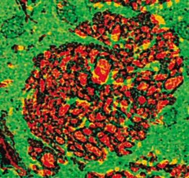
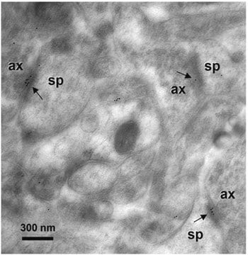
Anti-Glutamate Antibody detects level of Glutamate & has been published & validated for use in IH.
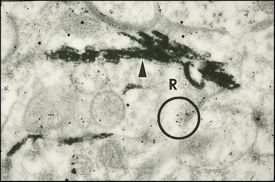
Immunohistochemistry: 1:2,500-1:10,000 using free floating sections by the PAP technique on rat hippocampus.
Optimal working dilutions must be determined by end user.
PROTOCOL for Glutamate Detection by Immunohisto/cytochemistry. Example for a rat brain.
1. SOLUTIONS TO BE PREPARED - Solution must be prepared as needed.
Solution A: 0.1M cacodylate, 10g/L sodium metabisulfite, pH 6.2.
Solution B: 0.1M cacodylate, 10g/L sodium metabisulfite, 3-5% glutaraldehyde, pH 7.5.
2. RAT PERFUSION - The rat is anaesthetized with sodium pentobarbital or Nembutal and perfused intracardially through the aorta using a pump with Solution A (30 mL): 150-300 mL/min, Solution B (500 mL): 150-300 mL/min.
3. POST FIXATION: 15 to 30 minutes in Solution B, then 4 soft washes in 0.05M Tris with 8.5 g/L sodium metabisulfite, pH 7.5 (Solution C) .
4. TISSUE SECTIONING: Vibratome or cryostat sections can be used.
5. REDUCTION STEP: Sections are reduced with Solution C containing 0.1M sodium borohydride for 10 minutes. The sections are washed 4 times in solution C without sodium borohydride.
6. APPLICATION OF GLUTAMATE ANTIBODY: Use a final dilution of 1:2,500-1:10,000 in Solution C containing 0.1% Triton X100 and 2% non-specific serum. Incubate 12 sections per 2 mL diluted antibody overnight, +2-8°C. Then wash the sections three times for 10 minutes each in Solution C. (Note that the antibody may be usable at a higher dilution. This should be explored to minimize the possibility of high background. Additionally, note that a change in the buffering system as indicated in the protocol may change the background and antibody recognition). The specific reaction is then revealed by PAP procedure.
7. SECOND ANTIBODY: Incubate the sections with a 1:50 to 1:200 dilution of goat anti-mouse in Solution B containing 1% non-specific serum for either 3 hrs at 20°C or 1-2 hr at 37°C. Then wash the sections, 3 times, for 10 minutes each with Solution C.
8. PAP: Incubate the sections with the appropriate dilution of peroxidase anti-peroxidase (for free floating method) in Solution C for 1-2 hours at 37°C. Then wash sections 3 times for 10 min each in solution C.
9. VISUALIZATION: The antigen-antibody complexes are visualized using DAB-4-HCl (25 mg/100 mL) (or other chromogen) in 0.05M Tris and filtrated; 0.05% hydrogen peroxide is added. Incubate the sections for 10 minutes at room temp. Stop the reaction by transferring the sections to 5 mL 0.05M Tris. Mount sections on chrome-alum coated slides. Dry overnight at 37°C. Rehydrate sections using conventional histological procedures. Coverslip using rapid mounting media.
For research use only; not for use as a diagnostic.
Research Category
Neuroscience
Neuroscience
Research Sub Category
Neurotransmitters & Receptors
Neuroinflammation & Pain
Neurotransmitters & Receptors
Neuroinflammation & Pain
Stato fisico
Format: Purified
Liquid in PBS containing 10mM sodium azide.
Stoccaggio e stabilità
Maintain at 2-8°C in undiluted aliquots for up to 6 months after date.
Note legali
CHEMICON is a registered trademark of Merck KGaA, Darmstadt, Germany
Esclusione di responsabilità
Unless otherwise stated in our catalog or other company documentation accompanying the product(s), our products are intended for research use only and are not to be used for any other purpose, which includes but is not limited to, unauthorized commercial uses, in vitro diagnostic uses, ex vivo or in vivo therapeutic uses or any type of consumption or application to humans or animals.
Non trovi il prodotto giusto?
Prova il nostro Motore di ricerca dei prodotti.
Codice della classe di stoccaggio
12 - Non Combustible Liquids
Classe di pericolosità dell'acqua (WGK)
WGK 2
Punto d’infiammabilità (°F)
Not applicable
Punto d’infiammabilità (°C)
Not applicable
Certificati d'analisi (COA)
Cerca il Certificati d'analisi (COA) digitando il numero di lotto/batch corrispondente. I numeri di lotto o di batch sono stampati sull'etichetta dei prodotti dopo la parola ‘Lotto’ o ‘Batch’.
Possiedi già questo prodotto?
I documenti relativi ai prodotti acquistati recentemente sono disponibili nell’Archivio dei documenti.
Monoclonal antibody directed against glutaraldehyde conjugated glutamate and immunocytochemical applications in the rat brain.
Chagnaud, J L, et al.
Brain Research, 481, 175-180 (1989)
Spatial learning and memory deficit of low level polybrominated diphenyl ethers-47 in male adult rat is modulated by intracellular glutamate receptors.
Tang Yan,Li Xiang,Jiang Xuejun,Chen Chengzhi,Qi Youbin,Yu Xuelan,Liu Yang,Peng Changyan,Chen Hui
The Journal of Toxicological Sciences null
Anatomical characterization of a rabbit cerebellar eyeblink premotor pathway using pseudorabies and identification of a local modulatory network in anterior interpositus.
Gonzalez-Joekes, J; Schreurs, BG
The Journal of Neuroscience null
Lin Cheng et al.
Cell research, 24(6), 665-679 (2014-03-19)
Neural progenitor cells (NPCs) can be induced from somatic cells by defined factors. Here we report that NPCs can be generated from mouse embryonic fibroblasts by a chemical cocktail, namely VCR (V, VPA, an inhibitor of HDACs; C, CHIR99021, an
Il team dei nostri ricercatori vanta grande esperienza in tutte le aree della ricerca quali Life Science, scienza dei materiali, sintesi chimica, cromatografia, discipline analitiche, ecc..
Contatta l'Assistenza Tecnica.