
推荐产品


材料
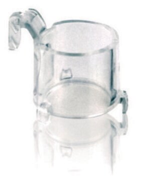
polystyrene housing
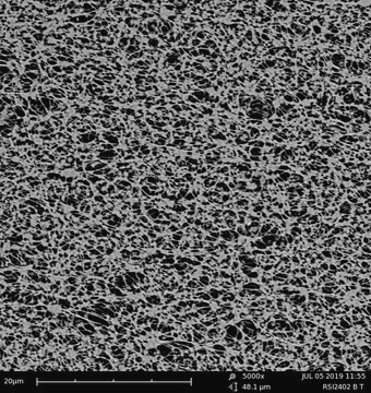
transparent PTFE membrane
品質等級
無菌
ethylene oxide treated
sterile
特點
hydrophilic
包裝
pack of 50 (individually blister packaged)
製造商/商標名
Millicell®
參數
50 °C max. temp.
技術
cell attachment: suitable
cell culture | mammalian: suitable
cell differentiation: suitable
高度
10.5 mm
直徑
12 mm
過濾面積
0.6 cm2
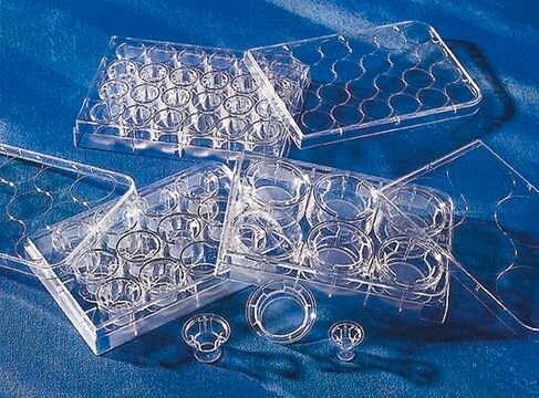
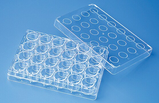
尺寸
24 wells
表面積
0.6 cm2
有效容積
0.6 mL
顏色
transparent membrane, when wetted
基質
Biopore™
孔徑
0.4 μm
結合類型
low binding surface
檢測方法
fluorometric
運輸包裝
ambient
相关类别
一般說明
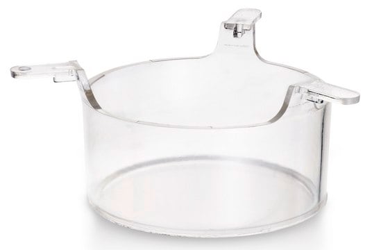
使用Millicell®嵌套板,附着或悬浮细胞可以从其上下两侧接触到培养基。更加密切地模拟了体内所发生的细胞生长、结构和功能。另外,Millicell®嵌套板使研究细胞单层的两侧成为可能。
Millicell®立式立式嵌套板:
-特别有助于细胞成长,为细胞研究提供了绝佳机会
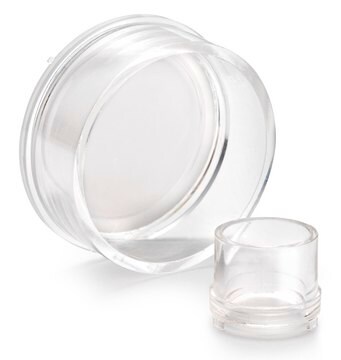
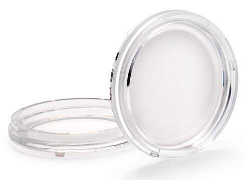
膜类型:
生物孔膜(亲水性 PTFE)
-用于低蛋白结合、活细胞观察和免疫荧光应用
应用
细胞附着、细胞生长、细胞分化、免疫细胞化学
應用
- Cell Attachment
- Cell Growth
- Cell Differentiation
- Immunocytochemistry
特點和優勢
- Promotes excellent cell growth and provides an exceptional opportunity for cell studies
- Low protein-binding hydrophilic PTFE membrane is ideal for live cell viewing and immunofluorescent application
法律資訊
推薦
相關產品
附屬品
儲存類別代碼
10-13 - German Storage Class 10 to 13
其他客户在看

实验方案
This is a Toluidine Blue Staining protocol listing materials and methods.
This is a Toluidine Blue Staining protocol listing materials and methods.
This protocol covers three methods for the microscopic examination of cell samples.
This protocol covers three methods for the microscopic examination of cell samples.
相关内容
This page covers the ECM coating protocols developed for four types of ECMs on Millicell®-CM inserts, Collagen Type 1, Fibronectin, Laminin, and Matrigel.
This page covers the ECM coating protocols developed for four types of ECMs on Millicell®-CM inserts, Collagen Type 1, Fibronectin, Laminin, and Matrigel.
我们的科学家团队拥有各种研究领域经验,包括生命科学、材料科学、化学合成、色谱、分析及许多其他领域.
联系技术服务部门