
MABN737
Anti-OPA1, clone 1OPA-1A8 Antibody
ascites fluid, clone 10PA-1A8, from mouse
别名:
Dynamin-like 120 kDa protein, mitochondrial, Large GTP-binding protein, LargeG, Optic atrophy protein 1 homolog, Dynamin-like 120 kDa protein, form S1
登录查看公司和协议定价
所有图片(2)
About This Item
分類程式碼代碼:
12352203
eCl@ss:
32160702
NACRES:
NA.41
推荐产品


生物源
mouse
品質等級
抗體表格
ascites fluid
抗體產品種類
primary antibodies
無性繁殖
10PA-1A8, monoclonal
物種活性
mouse, rat, human
技術
immunocytochemistry: suitable
immunoprecipitation (IP): suitable
western blot: suitable
同型
IgG1κ
NCBI登錄號
UniProt登錄號
運輸包裝
wet ice
目標翻譯後修改
unmodified
基因資訊
human ... OPA1(4976)
一般說明
OPA1 (optic atrophy 1) is a dynamin-related GTPase that plays a role in genome preservation and mitochondrial morphology. Localization of OPA1 is thought to be anchored to the inner membrane of the mitochondria. OPA1 is highly expressed in inner and outer retinal neurons and is important for normal mitochondrial function in those cells. Mutations in OPA1 are the major cause of autosomal dominant optic atrophy. Recent research has found that OPA1 also has the ability to protect neurons from excitotoxic injury and could be a potential clinical target to increase endurance of neurons post brain damage.
特異性
This antibody detects six known isoforms of OPA1 (Akepati, V. R., et al. (2008). J Neurochem. 106(1):372-383.).
免疫原
Recombinant protein corresponding to mouse OPA1.
應用
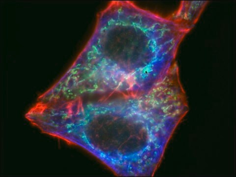
Immunocytochemistry Analysis: A 1:100 dilution from a representative lot detected OPA1 in COS cells (Prof. Mustapha Oulad-Abdelghani, IGBMC, France).
Immunoprecipitation Analysis: A representative lot from an independent laboratory immunoprecipitated OPA1 from mouse brain tissue lysate (Akepati, V. R., et al. (2008). J Neurochem. 106(1):372-383.).
Immunoprecipitation Analysis: A representative lot from an independent laboratory immunoprecipitated OPA1 from mouse brain tissue lysate (Akepati, V. R., et al. (2008). J Neurochem. 106(1):372-383.).
Research Category
Neuroscience
Neuroscience
Research Sub Category
Neurodegenerative Diseases
Neurodegenerative Diseases
This Anti-OPA1 antibody, clone 1OPA-1A8 is validated for use in western blotting, ICC & IP for the detection of OPA1.
品質
Evaluated by human brain tissue lysate.
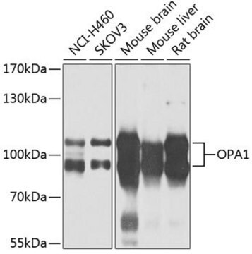
Western Blotting Analysis: A 1:1,000 dilution of this antibody detected OPA1 in 10 µg of human brain tissue lysate.
Western Blotting Analysis: A 1:1,000 dilution of this antibody detected OPA1 in 10 µg of human brain tissue lysate.
標靶描述
~90 and ~100 kDa observed. Six isoforms between 80 kDa and 100 kDa are known to exist (Akepati, V. R., et al. (2008). J Neurochem. 106(1):372-383.).
外觀
Unpurified
Mouse monoclonal IgG1κ ascites containing 0.05% sodium azide.
儲存和穩定性
Stable for 1 year at -20°C from date of receipt.
Handling Recommendations: Upon receipt and prior to removing the cap, centrifuge the vial and gently mix the solution. Aliquot into microcentrifuge tubes and store at -20°C. Avoid repeated freeze/thaw cycles, which may damage IgG and affect product performance.
Handling Recommendations: Upon receipt and prior to removing the cap, centrifuge the vial and gently mix the solution. Aliquot into microcentrifuge tubes and store at -20°C. Avoid repeated freeze/thaw cycles, which may damage IgG and affect product performance.
免責聲明
Unless otherwise stated in our catalog or other company documentation accompanying the product(s), our products are intended for research use only and are not to be used for any other purpose, which includes but is not limited to, unauthorized commercial uses, in vitro diagnostic uses, ex vivo or in vivo therapeutic uses or any type of consumption or application to humans or animals.
未找到合适的产品?
试试我们的产品选型工具.
儲存類別代碼
12 - Non Combustible Liquids
水污染物質分類(WGK)
nwg
閃點(°F)
Not applicable
閃點(°C)
Not applicable
Vasudheva Reddy Akepati et al.
Journal of neurochemistry, 106(1), 372-383 (2008-04-19)
OPA1, a nuclear encoded mitochondrial protein causing autosomal dominant optic atrophy, is a key player in mitochondrial fusion and cristae morphology regulation. In the present study, we have compared the OPA1 transcription and translation products of different mouse tissues. Unlike
我们的科学家团队拥有各种研究领域经验,包括生命科学、材料科学、化学合成、色谱、分析及许多其他领域.
联系技术服务部门