05-201
抗-Fas抗体(人,激活),克隆CH11
clone CH11, Upstate®, from mouse
别名:
APO-1 cell surface antigen, CD95 antigen, Fas (TNF receptor superfamily, member 6), Fas AMA, Fas antigen, apoptosis antigen 1, tumor necrosis factor receptor superfamily, member 6
登录查看公司和协议定价
所有图片(2)
About This Item
分類程式碼代碼:
12352203
eCl@ss:
32160702
NACRES:
NA.41
推荐产品
生物源
mouse
品質等級
抗體表格
affinity purified immunoglobulin
抗體產品種類
primary antibodies
無性繁殖
CH11, monoclonal
純化經由
affinity chromatography
物種活性
human
製造商/商標名
Upstate®
技術
flow cytometry: suitable
immunocytochemistry: suitable
western blot: suitable
同型
IgM
NCBI登錄號
UniProt登錄號
運輸包裝
dry ice
目標翻譯後修改
unmodified
基因資訊
human ... FAS(355)
一般說明
Fas/Apo-1/CD95(36 kDa)是肿瘤坏死因子(TNF)受体超家族(跨膜受体家族)的成员。Fas已被证明是凋亡细胞死亡的重要介质,并参与炎症。Fas配体(Fas-L)的结合诱导靶细胞膜中Fas的三聚化。Fas的激活通过Fas和FADD的死亡域之间的相互作用导致具有死亡域(FADD)的Fas相关蛋白募集。Procaspase 8通过FADD的死亡效应域(DED)与pro-caspase 8之间的相互作用与Fas结合的FADD结合,从而导致半胱天冬酶8的激活。活化的半胱天冬酶8裂解(激活)其他无活性的酶前体,实际上开始了半胱天冬酶级联反应,最终导致细胞凋亡。半胱天冬酶裂解核纤层蛋白,导致细胞核分解,失去正常结构。Fas诱导的细胞凋亡可以通过FLICE抑制蛋白(FLIP),Bcl-2或细胞因子反应修饰剂A(CrmA)在几个阶段有效阻断。
生物学活性
该抗体对表达Fas的人细胞具有细胞溶解活性。用编码人Fas的cDNA转染的小鼠WR19L细胞和L929细胞响应该抗体而发生细胞凋亡。
生物学活性
该抗体对表达Fas的人细胞具有细胞溶解活性。用编码人Fas的cDNA转染的小鼠WR19L细胞和L929细胞响应该抗体而发生细胞凋亡。
特異性
该抗体不识别TNF,也不与小鼠Fas发生交叉反应。 Fas配体可诱导人、小鼠和大鼠系统的细胞凋亡。
该抗体可识别在各种人细胞(包括髓样细胞、T淋巴母细胞和二倍体成纤维细胞)中表达的人细胞表面抗原Fas,Mr 43 kDa。
免疫原
FS-7(人二倍体成纤维细胞系)。 克隆CH-11。
應用
使用该抗Fas抗体(人,活化),克隆CH11检测Fas,已发表,并经验证可用于流式细胞术(FC)、免疫细胞化学(IC)和蛋白质印迹(WB)。
研究子类别
凋亡-附加
凋亡-附加
研究类别
细胞凋亡 & 癌症
细胞凋亡 & 癌症
蛋白质印迹:
0.5-2 μg/mL先前批次在Raji细胞裂解液中检测到Fas。
免疫细胞化学:
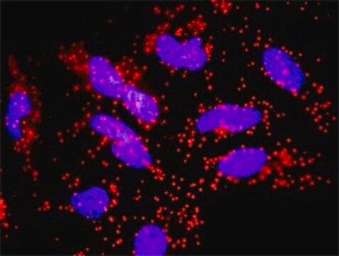
5-10 μg/mL的先前批次在用4%福尔马林/2%乙酸固定的HeLa细胞上,检测到Fas。
流式细胞术:
由独立实验室使用20 μg/mL的抗Fas克隆CH11测试先前批次的Fas(Yonehara, S., 1989; Kobayashi, N., 1990)。
0.5-2 μg/mL先前批次在Raji细胞裂解液中检测到Fas。
免疫细胞化学:
5-10 μg/mL的先前批次在用4%福尔马林/2%乙酸固定的HeLa细胞上,检测到Fas。
流式细胞术:
由独立实验室使用20 μg/mL的抗Fas克隆CH11测试先前批次的Fas(Yonehara, S., 1989; Kobayashi, N., 1990)。
品質
通过证明对表达Fas的人细胞的细胞溶解活性进行了常规评估。用编码人Fas的cDNA转染的小鼠WR19L细胞和L929细胞响应该抗体而发生细胞凋亡。
细胞凋亡分析:
15-20 µg/mL该批次最大程度地诱导人Jurkat细胞凋亡,处理24小时后死亡率为83%。
细胞凋亡分析:
15-20 µg/mL该批次最大程度地诱导人Jurkat细胞凋亡,处理24小时后死亡率为83%。
標靶描述
43 kDa
外觀
免疫亲和层析
纯化的小鼠单克隆IgM,溶于含有PBS(pH 7.2)和50%甘油的缓冲液中。 在-20ºC为液体形式。
儲存和穩定性
自收到之日起在-20°C可稳定保存1年。为了最大程度地回收产品,需在取下盖子之前将原始样品管进行离心。
分析報告
对照
人肝肿瘤、人乳腺肿瘤或Jurkat全细胞裂解液、Raji细胞裂解液。
人肝肿瘤、人乳腺肿瘤或Jurkat全细胞裂解液、Raji细胞裂解液。
其他說明
浓度:请参考批次特异性浓缩物的分析证书。
法律資訊
UPSTATE is a registered trademark of Merck KGaA, Darmstadt, Germany
免責聲明
除非我们的产品目录或产品附带的其他公司文档另有说明,否则我们的产品仅供研究使用,不得用于任何其他目的,包括但不限于未经授权的商业用途、体外诊断用途、离体或体内治疗用途或任何类型的消费或应用于人类或动物。
未找到合适的产品?
试试我们的产品选型工具.
儲存類別代碼
12 - Non Combustible Liquids
水污染物質分類(WGK)
WGK 2
閃點(°F)
Not applicable
閃點(°C)
Not applicable
G Linsinger et al.
Molecular and cellular biology, 19(5), 3299-3311 (1999-04-17)
Recent work suggests a participation of mitochondria in apoptotic cell death. This role includes the release of apoptogenic molecules into the cytosol preceding or after a loss of mitochondrial membrane potential DeltaPsim. The two uncouplers of oxidative phosphorylation carbonyl cyanide
F Basolo et al.
Laboratory investigation; a journal of technical methods and pathology, 80(9), 1413-1419 (2000-09-27)
The Fas-FasL system seems to mediate thyrocyte death in Hashimoto's thyroiditis. In thyroid cancer, down-regulation of bcl-2 seems to alter apoptosis control. We compared the expression of immunoreactive Fas and FasL in normal thyroid with that of tumors ranging from
M MacFarlane et al.
The Journal of cell biology, 148(6), 1239-1254 (2000-03-22)
Tumor necrosis factor-related apoptosis- inducing ligand (TRAIL) -induced apoptosis, in transformed human breast epithelial MCF-7 cells, resulted in a time-dependent activation of the initiator caspases-8 and -9 and the effector caspase-7. Cleavage of caspase-8 and its preferred substrate, Bid, preceded
A L Kim et al.
The Journal of biological chemistry, 274(49), 34924-34931 (1999-11-27)
A p53-derived C-terminal peptide induced rapid apoptosis in breast cancer cell lines carrying endogenous p53 mutations or overexpressed wild-type (wt) p53 but was not toxic to nonmalignant human cell lines containing wt p53. Apoptosis occurred through a Fas/APO-1 signaling pathway
J B Mannick et al.
Science (New York, N.Y.), 284(5414), 651-654 (1999-04-24)
Only a few intracellular S-nitrosylated proteins have been identified, and it is unknown if protein S-nitrosylation/denitrosylation is a component of signal transduction cascades. Caspase-3 zymogens were found to be S-nitrosylated on their catalytic-site cysteine in unstimulated human cell lines and
商品
Application note on how the CellASIC® ONIX2 microfluidic system can be used to analyze caspase-3 mediated apoptosis/cell death and cellular hypoxia in live immune and cancer cell lines.
我们的科学家团队拥有各种研究领域经验,包括生命科学、材料科学、化学合成、色谱、分析及许多其他领域.
联系技术服务部门


