Saltar al contenido
Merck
Todas las fotos(1)

Documentos clave

251M-1

Sigma-Aldrich

Factor XIIIa (AC-1A1) Mouse Monoclonal Antibody

Iniciar sesiónpara Ver la Fijación de precios por contrato y de la organización


About This Item

Código UNSPSC:
12352203
NACRES:
NA.41

origen biológico

mouse

Nivel de calidad

100
500

conjugado

unconjugated

forma del anticuerpo

diluted ascites fluid

tipo de anticuerpo

primary antibodies

clon

AC-1A1, monoclonal

descripción

For In Vitro Diagnostic Use in Select Regions (See Chart)

Formulario

buffered aqueous solution

reactividad de especies

human

envase

vial of 0.1 mL concentrate (251M-14)
vial of 0.5 mL concentrate (251M-15)
bottle of 1.0 mL predilute (251M-17)
vial of 1.0 mL concentrate (251M-16)
bottle of 7.0 mL predilute (251M-18)

fabricante / nombre comercial

Cell Marque

técnicas

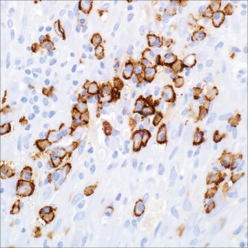
immunohistochemistry (formalin-fixed, paraffin-embedded sections): 1:100-1:500

isotipo

IgG1κ

control

dermatofibroma

Condiciones de envío

wet ice

temp. de almacenamiento

2-8°C

visualización

cytoplasmic

Información sobre el gen

human ... F13A1(2162)

Descripción general

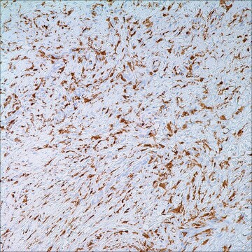
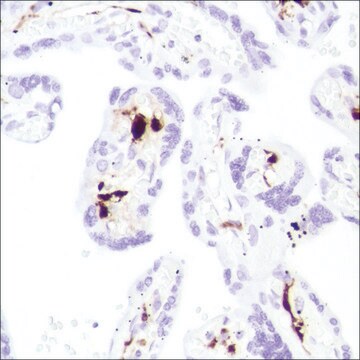
Factor XIIIa is a plasma transglutaminase that is part of the blood coagulation cascade and plays a critical role in stabilizing fibrin networks at the site of an injury to produce a clot. Factor XIIIa is expressed in platelets, megakaryocytes, fibroblast-like cells in the placenta, uterus, and prostate, monocytes and macrophages, and dermal dendritic cells. Anti-Factor XIIIa immunohistochemistry is used to identify fibrohistiocytic neoplasms such as dermatofibroma.

Factor XIIIa is a blood proenzyme that has been identified in platelets, megakaryocyte, and fibroblast-like mesenchymal or histiocytic cells present in the placenta, uterus, and prostate; it is also present in monocytes and macrophages and dermal dendritic cells. Anti- Factor XIIIa has been found to be useful in differentiating between dermatofibroma (90% (+)), dermatofibrosarcoma protuberans (25%(+)), and desmoplastic malignant melanoma (0%(+)). Factor XIIIa positivity is also seen in capillary hemagioblastoma (100%(+)), hemangioendothelioma (100%(+)), hemangiopericytoma (100%(+)), xanthogranuloma (100%(+)), xanthoma (100(+)), hepatocellular carcinoma (93%(+)), glomus tumor (80%(+)), and meningioma (80 % (+)).

Ligadura / enlace

Factor XIIIa Positive Control Slides , Product No. 251S, are available for immunohistochemistry (formalin-fixed, paraffin-embedded sections).

Forma física

Solution in Tris Buffer, pH 7.3-7.7, with 1% BSA and <0.1% Sodium Azide

Nota de preparación

Download the IFU specific to your product lot and formatNote: This requires a keycode which can be found on your packaging or product label.

Otras notas

For Technical Service please contact: 800-665-7284 or email: service@cellmarque.com

Información legal

Cell Marque is a trademark of Merck KGaA, Darmstadt, Germany

¿No encuentra el producto adecuado?  

Pruebe nuestro Herramienta de selección de productos.

Código de clase de almacenamiento

12 - Non Combustible Liquids

Clase de riesgo para el agua (WGK)

WGK 2

Punto de inflamabilidad (°F)

Not applicable

Punto de inflamabilidad (°C)

Not applicable


Elija entre una de las versiones más recientes:

Certificados de análisis (COA)

Lot/Batch Number

¿No ve la versión correcta?

Si necesita una versión concreta, puede buscar un certificado específico por el número de lote.

¿Ya tiene este producto?

Encuentre la documentación para los productos que ha comprado recientemente en la Biblioteca de documentos.

Visite la Librería de documentos

M G Horenstein et al.
The American journal of surgical pathology, 24(7), 996-1003 (2000-07-15)
Routine histology and immunohistochemistry can usually distinguish dermatofibroma (DF) and dermatofibrosarcoma protuberans (DFSP). DF generally expresses factor XIIIa whereas DFSP generally expresses CD34. The authors report 10 cutaneous fibrohistiocytic lesions combining clinical, histologic, and immunohistochemical features of both DF and
P Abenoza et al.
The American Journal of dermatopathology, 15(5), 429-434 (1993-10-01)
To assess the use of anti-CD34 and anti-factor-XIIIa antibodies for the differential diagnosis of dermatofibroma (DF) and dermatofibrosarcoma protuberans (DFSP), we stained 40 DFs and 13 DFSPs. A significant population of dendritic and spindle cells was reactive with anti-factor-XIIIa in
Louis P Dehner
The American journal of surgical pathology, 27(5), 579-593 (2003-04-30)
Juvenile xanthogranulomas (JXG) is a histiocytic disorder, primarily but not exclusively seen throughout the first two decades of life and principally as a solitary cutaneous lesion. This study is a retrospective clinical and pathologic review of 174 cases documenting the
A J Demetris et al.
The American journal of surgical pathology, 21(3), 263-270 (1997-03-01)
Epithelioid hemangioendothelioma (EHE) is best considered a vascular neoplasm of intermediate malignancy. Although usually progressive, the clinical course is highly unpredictable. The present communication describes a case of extensive recurrent hepatic EHE, limited to the liver allograft and initially manifest
E J Glusac et al.
The American journal of surgical pathology, 18(6), 583-590 (1994-06-01)
Epithelioid cell histiocytoma is a recently recognized lesion that is considered to be a variant of cutaneous fibrous histiocytoma (dermatofibroma). Ten cases are presented, including their light microscopic, immunohistochemical, and ultrastructural features. Eight of the cases are similar to those

Artículos

Immunohistochemistry (IHC) techniques and applications have greatly improved, dermatopathology is still largely based on H&E stained slides.This paper outlines ways in which IHC antibodies can be utilized for dermatopathology.

Nuestro equipo de científicos tiene experiencia en todas las áreas de investigación: Ciencias de la vida, Ciencia de los materiales, Síntesis química, Cromatografía, Analítica y muchas otras.

Póngase en contacto con el Servicio técnico