Saltar al contenido
Merck
Todas las fotos(4)

Key Documents

MABC32

Sigma-Aldrich

Anti-p62 (Sequestosome-1) Antibody, clone 11C9.2

clone 11C9.2, from mouse

Sinónimos:

sequestosome 1, EBI3-associated protein of 60 kDa, Paget disease of bone 3, phosphotyrosine independent ligand for the Lck SH2 domain p62, oxidative stress induced like, EBI3-associated protein p60, Phosphotyrosine-independent ligand for the Lck SH2 doma

Iniciar sesiónpara Ver la Fijación de precios por contrato y de la organización


About This Item

Código UNSPSC:
12352203
eCl@ss:
32160702
NACRES:
NA.41

origen biológico

mouse

Nivel de calidad

forma del anticuerpo

purified antibody

tipo de anticuerpo

primary antibodies

clon

11C9.2, monoclonal

reactividad de especies

mouse, rat, human

técnicas

flow cytometry: suitable
immunocytochemistry: suitable
western blot: suitable

isotipo

IgMκ

Nº de acceso NCBI

Nº de acceso UniProt

Condiciones de envío

wet ice

modificación del objetivo postraduccional

unmodified

Información sobre el gen

human ... SQSTM1(8878)

Descripción general

p62 is also known as sequestosome-1, and is a 62 kDa ras-GAP associated protein. p62 is thought to bind to src-family tyrosine kinases and may also provide a linker action for activated src-family kinases with their downstream effectors. p62 has an RNA-binding region and may have RNA-binding ability. p62 is associated with activation of transcription factor NF-κB, involved in cell differentiation, immune response and potassium channel regulation. p62 has recently been linked to extrinsic apoptosis and may be a key player in tumorigenesis.

Inmunógeno

GST-tagged recombinant protein corresponding to human p62 (Sequestosome-1).

Aplicación

Detect p62 (Sequestosome-1) using this Anti-p62 (Sequestosome-1) Antibody, clone 11C9.2 validated for use in WB, FC & IC.
Research Category
Apoptosis & Cancer
Research Sub Category
Apoptosis - Additional
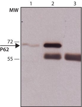
Western Blot Analysis: 0.001 µg/mL from a previous lot detected p62 (Sequestosome-1) in 10 µg of PC12 cell lysate.

Flow Cytometry Analysis: 1.0 µg from a previous lot detected p62 (Sequestosome-1) in the staining of fixed and permeabilized HeLa cells.

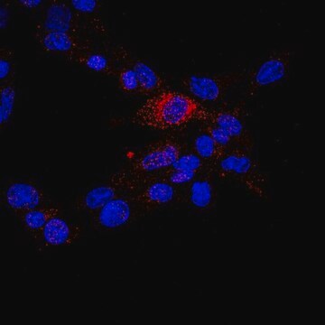
Immunocytochemistry Analysis: A 1:500 dilution from a previous lot detected p62 (Sequestosome-1) in NIH/3T3, A431, and HeLa cells.

Calidad

Evaluated by Western Blot in A431 cell lysate.

Western Blot Analysis: 0.001 µg/mL of this antibody detected p62 (Sequestosome-1) in 10 µg of A431 cell lysate.

Descripción de destino

~47-60 kDa observed.
The calculated molecular weight of this protein is 47 kDa and also has an isoform at 38 kDa Due to modifications, this protein may be observed up to ~60 kDa in some lysates.

Forma física

Format: Purified
Purified mouse monoclonal IgMκ in buffer containing PBS with 0.05% sodium azide.

Almacenamiento y estabilidad

Stable for 1 year at 2-8°C from date of receipt.

Nota de análisis

Control
A431 cell lysate

Cláusula de descargo de responsabilidad

Unless otherwise stated in our catalog or other company documentation accompanying the product(s), our products are intended for research use only and are not to be used for any other purpose, which includes but is not limited to, unauthorized commercial uses, in vitro diagnostic uses, ex vivo or in vivo therapeutic uses or any type of consumption or application to humans or animals.

¿No encuentra el producto adecuado?  

Pruebe nuestro Herramienta de selección de productos.

Opcional

Referencia del producto
Descripción
Precios

Código de clase de almacenamiento

12 - Non Combustible Liquids

Clase de riesgo para el agua (WGK)

WGK 2

Punto de inflamabilidad (°F)

Not applicable

Punto de inflamabilidad (°C)

Not applicable


Certificados de análisis (COA)

Busque Certificados de análisis (COA) introduciendo el número de lote del producto. Los números de lote se encuentran en la etiqueta del producto después de las palabras «Lot» o «Batch»

¿Ya tiene este producto?

Encuentre la documentación para los productos que ha comprado recientemente en la Biblioteca de documentos.

Visite la Librería de documentos

AMPK involvement in endoplasmic reticulum stress and autophagy modulation after fatty liver graft preservation: a role for melatonin and trimetazidine cocktail.
Zaouali, Mohamed Amine, et al.
Journal of Pineal Research (2013)
Huimin Yan et al.
Journal of rheumatic diseases and treatment, 1(1), 5-5 (2015-06-30)
Autophagy plays a central role in various disease processes. However, its contribution to inflammatory arthritides such as rheumatoid arthritis (RA) is unclear. We observed that autophagy is engaged in the K/BxN serum transfer model of RA but autophagic flux is
Michalis Papadakis et al.
Nature medicine, 19(3), 351-357 (2013-02-26)
Previous attempts to identify neuroprotective targets by studying the ischemic cascade and devising ways to suppress it have failed to translate to efficacious therapies for acute ischemic stroke. We hypothesized that studying the molecular determinants of endogenous neuroprotection in two
Mallika Valapala et al.
Autophagy, 10(3), 480-496 (2014-01-29)
In phagocytic cells, including the retinal pigment epithelium (RPE), acidic compartments of the endolysosomal system are regulators of both phagocytosis and autophagy, thereby helping to maintain cellular homeostasis. The acidification of the endolysosomal system is modulated by a proton pump
Elisa Teyssou et al.
Neurobiology of aging, 58, 239-239 (2017-07-19)
Mutations in UBQLN2 have been associated with rare cases of X-linked juvenile and adult forms of amyotrophic lateral sclerosis (ALS) and ALS linked to frontotemporal dementia (FTD). Here, we report 1 known (c.1489C>T, p.Pro497Ser, P497S) and 3 novel (c.1481C>T, p.Pro494Leu

Artículos

Autophagy is a highly regulated process that is involved in cell growth, development, and death. In autophagy cells destroy their own cytoplasmic components in a very systematic manner and recycle them.

Nuestro equipo de científicos tiene experiencia en todas las áreas de investigación: Ciencias de la vida, Ciencia de los materiales, Síntesis química, Cromatografía, Analítica y muchas otras.

Póngase en contacto con el Servicio técnico