Direkt zum Inhalt
Merck
Alle Fotos(3)

Key Documents

MABS1266

Sigma-Aldrich

Anti-LOX-1 Antibody, clone 15C4

clone 15C4, from mouse

Synonym(e):

Oxidized low-density lipoprotein receptor 1, Ox-LDL receptor 1, C-type lectin domain family 8 member A, Lectin-like oxidized LDL receptor 1, Lectin-like oxLDL receptor 1, hLOX-1, Lectin-type oxidized LDL receptor 1

Anmeldenzur Ansicht organisationsspezifischer und vertraglich vereinbarter Preise


About This Item

UNSPSC-Code:
12352203
eCl@ss:
32160702

Biologische Quelle

mouse

Qualitätsniveau

Antikörperform

purified immunoglobulin

Antikörper-Produkttyp

primary antibodies

Klon

15C4, monoclonal

Speziesreaktivität

human

Methode(n)

flow cytometry: suitable
immunofluorescence: suitable
immunohistochemistry: suitable
western blot: suitable

Isotyp

IgG2aκ

NCBI-Hinterlegungsnummer

UniProt-Hinterlegungsnummer

Versandbedingung

ambient

Posttranslationale Modifikation Target

unmodified

Angaben zum Gen

human ... OLR1(4973)

Allgemeine Beschreibung

Oxidized low-density lipoprotein receptor 1 (UniProt: P78380; also known as Ox-LDL receptor 1, C-type lectin domain family 8 member A, Lectin-like oxidized LDL receptor 1, LOX-1, Lectin-like oxLDL receptor 1, hLOX-1, Lectin-type oxidized LDL receptor 1) is encoded by the OLR1 (also known as CLEC8A, LOX1) gene (Gene ID: 4973) in human. LOX1 is a type II transmembrane receptor for oxidatively modified low density lipoprotein (oxLDL), a well-known marker of atherosclerosis. A free form of LOX-1 has also been reported. LOX-1 has a The C-type lectin domain that mediates the recognition and binding of oxLDL. Its cytoplasmic region is considered to be essential for subcellular sorting on the cell surface. LOX-1 is expressed at high level in endothelial cells and vascular-rich organs such as placenta, lung, liver and brain, aortic intima, bone marrow, spinal cord and substantia nigra. It is also expressed at the surface of dendritic cells. LOX-1 mediates the recognition, internalization and degradation of oxLDL by vascular endothelial cells. OxLDL induces vascular endothelial cell activation and dysfunction, resulting in pro-inflammatory responses, pro-oxidative conditions and also apoptosis. In addition to binding oxLDL, LOX-1 also acts as a receptor for the HSP70 protein involved in antigen cross-presentation to naive T-cells in dendritic cells, thereby participating in cell-mediated antigen cross-presentation. LOX-1 is up-regulated in atherosclerotic lesions, by oxLDL, reactive oxygen species and fluid shear stress.

Spezifität

Clone 15C4 recognizes LOX-1 in human cells.

Immunogen

Human LOX 1 ectodomain-hIgGFc fusion protein.

Anwendung

Research Category
Zelluläre Signaltransduktion
Detect LOX-1 using this mouse monoclonal Anti-LOX-1 Antibody, clone 15C4, Cat. No. MABS1266. It has been tested in Cell Differentiation studies, Flow Cytometry, Immunofluorescence, Immunohistochemistry, and Western Blotting.
Immunohistochemistry Analysis: A 1:250-1,000 dilution from a representative lot detected LOX-1 in human spleen, human large intestine, and human vagina tissues.

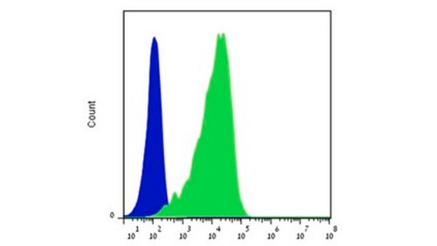
Flow Cytometry Analysis: A representative lot detected LOX-1 in Flow Cytometry applications (Li, D., et. al. (2012). J Exp Med. 209(1):109-21).

Immunofluorescence Analysis: A representative lot detected LOX-1 in Immunofluorescence applications (Li, D., et. al. (2012). J Exp Med. 209(1):109-21).

Cell Differentiation Analysis: A representative lot detected LOX-1 in Cell Differentiation applications (Joo, H., et. al. (2014). Immunity. 41(4):592-604).

Immunohistochemistry Analysis: A representative lot detected LOX-1 in Immunohistochemistry applications (Duluc, D., et. al. (2013). Microb Pathog. 58:35-44).

Flow Cytometry Analysis: 1 ug from a representative lot detected LOX-1 in one million A549 cells.

Qualität

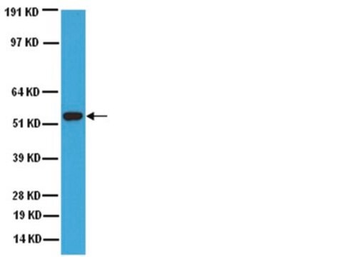
Evaluated by Western Blotting in human liver tissue lysates.

Western Blotting Analysis: 2 µg/mL of this antibody detected LOX-1 in 10 µg of human liver tissue lysates.

Zielbeschreibung

30.96 kDa calculated. Uncharacterized band(s) may appear in some lysates.

Physikalische Form

Protein G purified
Format: Purified
Purified mouse monoclonal antibody IgG2a in PBS without preservatives.

Lagerung und Haltbarkeit

Stable for 1 year at -20°C from date of receipt.

Handling Recommendations: Upon receipt and prior to removing the cap, centrifuge the vial and gently mix the solution. Aliquot into microcentrifuge tubes and store at -20°C. Avoid repeated freeze/thaw cycles, which may damage IgG and affect product performance.

Sonstige Hinweise

Concentration: Please refer to lot specific datasheet.

Haftungsausschluss

Unless otherwise stated in our catalog or other company documentation accompanying the product(s), our products are intended for research use only and are not to be used for any other purpose, which includes but is not limited to, unauthorized commercial uses, in vitro diagnostic uses, ex vivo or in vivo therapeutic uses or any type of consumption or application to humans or animals.

Sie haben nicht das passende Produkt gefunden?  

Probieren Sie unser Produkt-Auswahlhilfe. aus.

Lagerklassenschlüssel

12 - Non Combustible Liquids

WGK

WGK 2

Flammpunkt (°F)

Not applicable

Flammpunkt (°C)

Not applicable


Analysenzertifikate (COA)

Suchen Sie nach Analysenzertifikate (COA), indem Sie die Lot-/Chargennummer des Produkts eingeben. Lot- und Chargennummern sind auf dem Produktetikett hinter den Wörtern ‘Lot’ oder ‘Batch’ (Lot oder Charge) zu finden.

Besitzen Sie dieses Produkt bereits?

In der Dokumentenbibliothek finden Sie die Dokumentation zu den Produkten, die Sie kürzlich erworben haben.

Die Dokumentenbibliothek aufrufen

Unser Team von Wissenschaftlern verfügt über Erfahrung in allen Forschungsbereichen einschließlich Life Science, Materialwissenschaften, chemischer Synthese, Chromatographie, Analytik und vielen mehr..

Setzen Sie sich mit dem technischen Dienst in Verbindung.