Passa al contenuto
Merck
Tutte le immagini(4)

Documenti fondamentali

P3555

Sigma-Aldrich

Monoclonal Anti-Phosphothreonine antibody produced in mouse

clone PTR-8, ascites fluid

Sinonimo/i:

Monoclonal Anti-Phosphothreonine, Phospho Thr, Phospho Threonine, Phospho−Thr, Phospho-Threonine, p-Thr

Autenticatiper visualizzare i prezzi riservati alla tua organizzazione & contrattuali


About This Item

Codice UNSPSC:
12352203
NACRES:
NA.44

Origine biologica

mouse

Livello qualitativo

Coniugato

unconjugated

Forma dell’anticorpo

ascites fluid

Tipo di anticorpo

primary antibodies

Clone

PTR-8, monoclonal

contiene

15 mM sodium azide

tecniche

indirect ELISA: 1:4,000
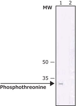
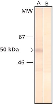
western blot: 1:50

Isotipo

IgG2b

Condizioni di spedizione

dry ice

Temperatura di conservazione

−20°C

modifica post-traduzionali bersaglio

unmodified

Cerchi prodotti simili? Visita Guida al confronto tra prodotti

Descrizione generale

As determined by ELISA and dot blot, the antibody reacts specifically with phosphorylated threonine, both as free amino acid or conjugated to carriers such as BSA or KLH. No cross-reactivity is observed with non-phosphorylated threonine, phosphoserine, phosphotyrosine, AMP or ATP. This antibody has been used in immunoblotting for the localization of some phosphothreonine-containing proteins. Certain proteins known to contain phosphorylated threonine may not be recognized by this antibody due to steric hindrance of the recognition site.
Monoclonal Anti-Phosphothreonine (mouse IgG2b isotype) is derived from the hybridoma produced by the fusion of mouse myeloma cells and splenocytes from an immunized mouse.

Immunogeno

phosphothreonine conjugated to keyhole limpet hemocyanin (KLH).

Applicazioni

Monoclonal Anti-Phosphothreonine antibody produced in mouse has been used in:immunoblotting, enzyme linked immuno sorbent assay (ELISA) ,dot blot.
Monoclonal and polyclonal antibodies directed against phosphorylated residues may be useful as analytical and preparative tools, by enabling the identification, quantification and immunoaffinity isolation of phosphorylated cellular proteins. Antibodies can be employed to monitor alterations in phosphorylation of specific proteins as they occur in intact organs or even whole animals.
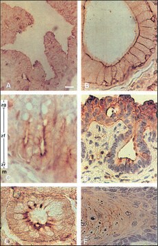
Mouse monoclonal clone PTR-8 anti-phosphothreonine antibody may be used for the localization of phosphorylated threonine using various immunochemical assays such as ELISA, dot blot, and immunoblotting. Due to steric hindrance of the recognition site, this antibody may not recognize certain proteins known to contain phosphorylated threonine.
Mouse monoclonal clone PTR-8 anti-Phosphothreonine antibody reacts with phosphorylated threonine both as a free amino acid or when conjugated to carriers such as BSA or KLH, using ELISA and dot blot. It does not react with nonphosphorylated threonine, phosphorylated tyrosine or serine, AMP or ATP.

Azioni biochim/fisiol

Protein phosphorylation and dephosphorylation are basic mechanisms for the modification of protein function in eukaryotic cells. Phosphorylation is a rare post-translational event in normal tissue. However, the abundance of phosphorylated cellular proteins increases tenfold following various activation processes, which are mediated through phosphotyrosine, phosphoserine or phosphothreonine (p-Tyr/p-Ser/p-Thr). Many different mitogenic systems, such as the EGF, PDGF and insulin receptor systems, contain Tyr/Ser/Thr kinase domains that autophosphorylate specific Tyr/Ser/Thr residues upon binding of their ligands. T cell antigen receptor complex or receptors for some hemopoietic growth factors may stimulate associated kinases, and cells transformed by viral oncogenes contain elevated levels of phosphorylated Tyr/Ser/Thr. An understanding of transformation by oncogenes and mitogenic processes of growth factors depends on the identification of their substrate and a subsequent determination of how phosphorylation affects the properties of these proteins.

Esclusione di responsabilità

Unless otherwise stated in our catalog or other company documentation accompanying the product(s), our products are intended for research use only and are not to be used for any other purpose, which includes but is not limited to, unauthorized commercial uses, in vitro diagnostic uses, ex vivo or in vivo therapeutic uses or any type of consumption or application to humans or animals.

Non trovi il prodotto giusto?  

Prova il nostro Motore di ricerca dei prodotti.

Prodotti correlati

N° Catalogo
Descrizione
Determinazione del prezzo

Codice della classe di stoccaggio

10 - Combustible liquids

Classe di pericolosità dell'acqua (WGK)

WGK 3

Punto d’infiammabilità (°F)

Not applicable

Punto d’infiammabilità (°C)

Not applicable


Scegli una delle versioni più recenti:

Certificati d'analisi (COA)

Lot/Batch Number

Non trovi la versione di tuo interesse?

Se hai bisogno di una versione specifica, puoi cercare il certificato tramite il numero di lotto.

Possiedi già questo prodotto?

I documenti relativi ai prodotti acquistati recentemente sono disponibili nell’Archivio dei documenti.

Visita l’Archivio dei documenti

Clémence Rougeaux et al.
Cellular microbiology, 10(3), 632-654 (2007-11-06)
Human decay accelerating factor (hDAF, CD55) and members of the carcinoembryonic-antigen-related cell-adhesion molecules (hCEACAMs) family are recognized as receptors by Gram-negative, diffusely adhering Escherichia coli (DAEC) strains expressing Afa/Dr adhesins. We report here that hCEACAM1-4L has a key function in
Quantification of the Dynamic Phosphorylation Process Of ERK Using Stable Isotope Dilution Selective Reaction Monitoring Mass Spectrometry
Lee N, et al.
Proteomics, 1900086-1900086 (2019)
Takeru Zama et al.
The Journal of biological chemistry, 277(26), 23909-23918 (2002-04-18)
Mitogen-activated protein kinases (MAPKs) are activated in response to various extracellular stimuli, and their activities are regulated by upstream activating kinases and protein phosphatases such as MAPK phosphatases (MKPs). We report the identification and characterization of a novel MKP termed
Min-Yeon Choi et al.
Molecules and cells, 26(2), 200-205 (2008-07-04)
The mitogen-activated protein kinase (MAPK) signaling pathway is activated in response to extracellular stimuli and regulates various activities in eukaryotic cells. Following exposure to stimuli, MAPK is known to be activated via dual phosphorylation at a conserved TxY motif in
Carsten Tue Berg et al.
Journal of neuroinflammation, 14(1), 127-127 (2017-06-26)
Antibodies with specificity for myelin oligodendrocyte glycoprotein (MOG) are implicated in multiple sclerosis and related diseases. The pathogenic importance of anti-MOG antibody in primary demyelinating pathology remains poorly characterized. The objective of this study is to investigate whether administration of

Il team dei nostri ricercatori vanta grande esperienza in tutte le aree della ricerca quali Life Science, scienza dei materiali, sintesi chimica, cromatografia, discipline analitiche, ecc..

Contatta l'Assistenza Tecnica.