Passa al contenuto
Merck
Tutte le immagini(10)

Documenti fondamentali

HPA008461

Sigma-Aldrich

ANTI-SUN1 antibody produced in rabbit

enhanced validation

Ab2, Prestige Antibodies® Powered by Atlas Antibodies, affinity isolated antibody, buffered aqueous glycerol solution

Sinonimo/i:

Anti-Sad1/unc-84 protein-like 1, Anti-UNC84A, Anti-Unc-84 homolog A

Autenticatiper visualizzare i prezzi riservati alla tua organizzazione & contrattuali


About This Item

Codice UNSPSC:
12352203
Numero Human Protein Atlas:
NACRES:
NA.43

Origine biologica

rabbit

Livello qualitativo

Coniugato

unconjugated

Forma dell’anticorpo

affinity isolated antibody

Tipo di anticorpo

primary antibodies

Clone

polyclonal

Nome Commerciale

Prestige Antibodies® Powered by Atlas Antibodies

Stato

buffered aqueous glycerol solution

Reattività contro le specie

human

Convalida avanzata

independent
Learn more about Antibody Enhanced Validation

tecniche

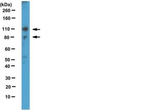
immunoblotting: 0.04-0.4 μg/mL
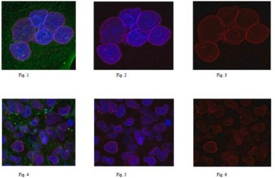
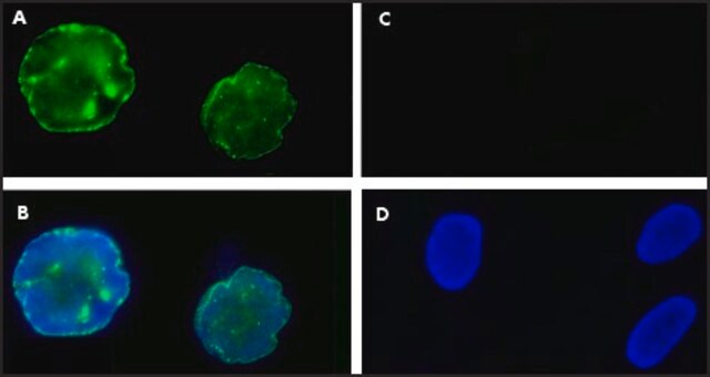
immunofluorescence: 0.25-2 μg/mL
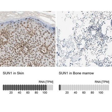
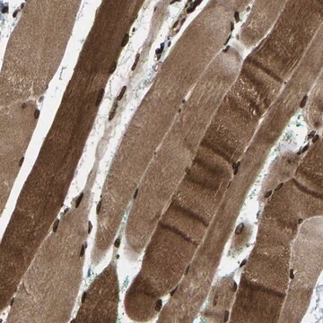
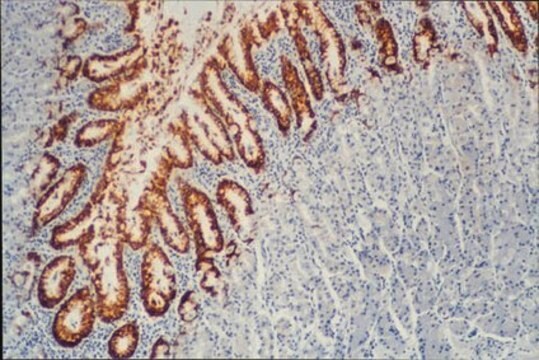
immunohistochemistry: 1:200-1:500

Sequenza immunogenica

QGNFFSFLPVLNWASMHRTQRVDDPQDVFKPTTSRLKQPLQGDSEAFPWHWMSGVEQQVASLSGQCHHHGENLRELTTLLQKLQARVDQMEGGAAGPSASVRDAVGQPPRETDFMAFHQEHEVRMSHLEDILGKLR

Condizioni di spedizione

wet ice

Temperatura di conservazione

−20°C

modifica post-traduzionali bersaglio

unmodified

Informazioni sul gene

human ... UNC84A(23353)

Cerchi prodotti simili? Visita Guida al confronto tra prodotti

Descrizione generale

Sad1 and UNC84 domain containing 1 (SUN1) is present in the inner nuclear envelope. It has a C-terminal domain called the SUN domain that is located in the space between the inner and outer nuclear membranes and an N-terminal domain located in the nucleoplasm.

Immunogeno

Sad1/unc-84 protein-like 1 recombinant protein epitope signature tag (PrEST)

Applicazioni

Anti-SUN1 antibody produced in rabbit, a Prestige Antibody, is developed and validated by the Human Protein Atlas (HPA) project . Each antibody is tested by immunohistochemistry against hundreds of normal and disease tissues. These images can be viewed on the Human Protein Atlas (HPA) site by clicking on the Image Gallery link. The antibodies are also tested using immunofluorescence and western blotting. To view these protocols and other useful information about Prestige Antibodies and the HPA, visit sigma.com/prestige. Anti-SUN1 antibody produced in rabbit is also suitable for fluorescent staining.
Applications in which this antibody has been used successfully, and the associated peer-reviewed papers, are given below.
Immunofluorescence (1 paper)

Azioni biochim/fisiol

Sad1 and UNC84 domain containing 1 (SUN1) plays an important role in connecting telomeres with the nuclear envelope. It binds to telomere-binding proteins and helps in the formation of the meiotic bouquet. This is a special cluster formed by telomeres at the nuclear envelope and occurs before meiosis I. SUN1 also interacts with SUN2 to form homodimers and heterodimers. This in turn helps in aiding the interaction between the nuclear envelope and the centrosome. It has also been shown that reforming nuclear envelope employs SUN1 to acetylate histones for de-condensation of DNA after mitosis. It is one of the earliest factors to associate with segregated daughter chromosomes in anaphase.

Caratteristiche e vantaggi

Prestige Antibodies® are highly characterized and extensively validated antibodies with the added benefit of all available characterization data for each target being accessible via the Human Protein Atlas portal linked just below the product name at the top of this page. The uniqueness and low cross-reactivity of the Prestige Antibodies® to other proteins are due to a thorough selection of antigen regions, affinity purification, and stringent selection. Prestige antigen controls are available for every corresponding Prestige Antibody and can be found in the linkage section.

Every Prestige Antibody is tested in the following ways:
  • IHC tissue array of 44 normal human tissues and 20 of the most common cancer type tissues.
  • Protein array of 364 human recombinant protein fragments.

Linkage

Corresponding Antigen APREST71107

Stato fisico

Solution in phosphate-buffered saline, pH 7.2, containing 40% glycerol and 0.02% sodium azide

Note legali

Prestige Antibodies is a registered trademark of Merck KGaA, Darmstadt, Germany

Esclusione di responsabilità

Unless otherwise stated in our catalog or other company documentation accompanying the product(s), our products are intended for research use only and are not to be used for any other purpose, which includes but is not limited to, unauthorized commercial uses, in vitro diagnostic uses, ex vivo or in vivo therapeutic uses or any type of consumption or application to humans or animals.

Non trovi il prodotto giusto?  

Prova il nostro Motore di ricerca dei prodotti.

Codice della classe di stoccaggio

10 - Combustible liquids

Classe di pericolosità dell'acqua (WGK)

WGK 1

Punto d’infiammabilità (°F)

Not applicable

Punto d’infiammabilità (°C)

Not applicable

Dispositivi di protezione individuale

Eyeshields, Gloves, multi-purpose combination respirator cartridge (US)


Scegli una delle versioni più recenti:

Certificati d'analisi (COA)

Lot/Batch Number

Non trovi la versione di tuo interesse?

Se hai bisogno di una versione specifica, puoi cercare il certificato tramite il numero di lotto.

Possiedi già questo prodotto?

I documenti relativi ai prodotti acquistati recentemente sono disponibili nell’Archivio dei documenti.

Visita l’Archivio dei documenti

Toshiro Anno et al.
Biochemical and biophysical research communications, 424(1), 94-99 (2012-06-26)
The linker of nucleus and cytoskeleton (LINC) complex, including nesprin-1, has been suggested to be crucial for many biological processes. Previous studies have shown that mutations in nesprin-1 cause abnormal cellular functions and diseases, possibly because of insufficient force transmission
Jennifer M Bupp et al.
The Journal of cell biology, 179(5), 845-854 (2007-11-28)
Positioning of telomeres at the nuclear periphery can have dramatic effects on gene expression by establishment of heritable, transcriptionally repressive subdomains. However, little is known about the integral membrane proteins that mediate telomere tethering at the nuclear envelope. Here, we
Ya-Hui Chi et al.
The Journal of biological chemistry, 282(37), 27447-27458 (2007-07-17)
Replicated mammalian chromosomes condense to segregate during anaphase, and they de-condense at the conclusion of mitosis. Currently, it is not understood what the factors and events are that specify de-condensation. Here, we demonstrate that chromosome de-condensation needs the function of
Qiang Wang et al.
DNA and cell biology, 25(10), 554-562 (2006-11-30)
The nuclear envelope forms a selective barrier that separates the cytoplasm from the nucleus. During mitosis the nuclear envelope breaks down so that the microtubule network can form contacts with the kinetochore and guide chromosome segregation. Previous studies have suggested
Hamish T J Gilbert et al.
Nature communications, 10(1), 4149-4149 (2019-09-14)
Studies of cellular mechano-signaling have often utilized static models that do not fully replicate the dynamics of living tissues. Here, we examine the time-dependent response of primary human mesenchymal stem cells (hMSCs) to cyclic tensile strain (CTS). At low-intensity strain

Il team dei nostri ricercatori vanta grande esperienza in tutte le aree della ricerca quali Life Science, scienza dei materiali, sintesi chimica, cromatografia, discipline analitiche, ecc..

Contatta l'Assistenza Tecnica.