Passa al contenuto
Merck
Tutte le immagini(6)

Documenti fondamentali

AMAB90556

Sigma-Aldrich

Monoclonal Anti-NES antibody produced in mouse

enhanced validation

Prestige Antibodies® Powered by Atlas Antibodies, clone CL0197, purified immunoglobulin, buffered aqueous glycerol solution

Sinonimo/i:

FLJ21841

Autenticatiper visualizzare i prezzi riservati alla tua organizzazione & contrattuali


About This Item

Codice UNSPSC:
12352203
NACRES:
NA.41
Coniugato:
unconjugated
application:
IF
IHC
Clone:
CL0197, monoclonal
Reattività contro le specie:
human
citations:
15
tecniche:
immunoblotting: 1 μg/mL
immunofluorescence: 2-10 μg/mL (Fixation/Permeabilization: PFA/Triton X-100)
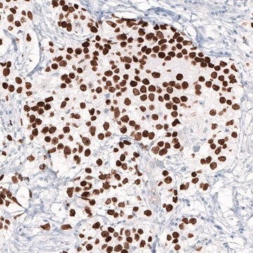
immunohistochemistry: 1:2500- 1:5000

Origine biologica

mouse

Livello qualitativo

Coniugato

unconjugated

Forma dell’anticorpo

purified immunoglobulin

Tipo di anticorpo

primary antibodies

Clone

CL0197, monoclonal

Nome Commerciale

Prestige Antibodies® Powered by Atlas Antibodies

Stato

buffered aqueous glycerol solution

Reattività contro le specie

human

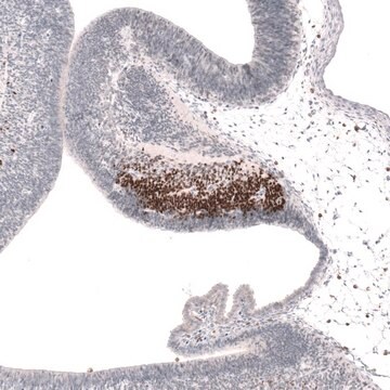
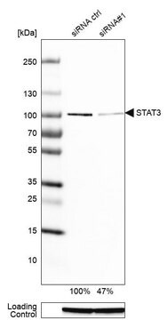
Convalida avanzata

RNAi knockdown
Learn more about Antibody Enhanced Validation

tecniche

immunoblotting: 1 μg/mL
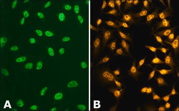
immunofluorescence: 2-10 μg/mL (Fixation/Permeabilization: PFA/Triton X-100)
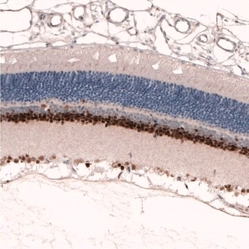
immunohistochemistry: 1:2500- 1:5000

Isotipo

IgG1

N° accesso Ensembl | uomo

N° accesso UniProt

Condizioni di spedizione

wet ice

Temperatura di conservazione

−20°C

modifica post-traduzionali bersaglio

unmodified

Informazioni sul gene

human ... NES(10763)

Immunogeno

nestin recombinant protein epitope signature tag (PrEST)

Sequence
DPEGQSQQVGAPGLQAPQGLPEAIEPLVEDDVAPGGDQASPEVMLGSEPAMGESAAGAEPGPGQGVGGLGDPGHLTREEVMEPPLEEESLEAKRVQGLEGPRKDLEEAGGLGTEFSELP

Epitope
Binds to an epitope located within the peptide sequence VGGLGDPGHL as determined by overlapping synthetic peptides.

Applicazioni

All Prestige Antibodies Powered by Atlas Antibodies are developed and validated by the Human Protein Atlas (HPA) project and as a result, are supported by the most extensive characterization in the industry.

The Human Protein Atlas project can be subdivided into three efforts: Human Tissue Atlas, Cancer Atlas, and Human Cell Atlas. The antibodies that have been generated in support of the Tissue and Cancer Atlas projects have been tested by immunohistochemistry against hundreds of normal and disease tissues and through the recent efforts of the Human Cell Atlas project, many have been characterized by immunofluorescence to map the human proteome not only at the tissue level but now at the subcellular level. These images and the collection of this vast data set can be viewed on the Human Protein Atlas (HPA) site by clicking on the Image Gallery link. We also provide Prestige Antibodies® protocols and other useful information.

Caratteristiche e vantaggi

Prestige Antibodies® are highly characterized and extensively validated antibodies with the added benefit of all available characterization data for each target being accessible via the Human Protein Atlas portal linked just below the product name at the top of this page. The uniqueness and low cross-reactivity of the Prestige Antibodies® to other proteins are due to a thorough selection of antigen regions, affinity purification, and stringent selection. Prestige antigen controls are available for every corresponding Prestige Antibody and can be found in the linkage section.

Every Prestige Antibody is tested in the following ways:
  • IHC tissue array of 44 normal human tissues and 20 of the most common cancer type tissues.
  • Protein array of 364 human recombinant protein fragments.

Linkage

Corresponding Antigen APREST70691

Stato fisico

Phospate buffered saline, pH 7.2, containing 40% glycerol and 0.02% sodium azide

Note legali

Prestige Antibodies is a registered trademark of Merck KGaA, Darmstadt, Germany

Esclusione di responsabilità

Unless otherwise stated in our catalog or other company documentation accompanying the product(s), our products are intended for research use only and are not to be used for any other purpose, which includes but is not limited to, unauthorized commercial uses, in vitro diagnostic uses, ex vivo or in vivo therapeutic uses or any type of consumption or application to humans or animals.

Non trovi il prodotto giusto?  

Prova il nostro Motore di ricerca dei prodotti.

Codice della classe di stoccaggio

10 - Combustible liquids

Classe di pericolosità dell'acqua (WGK)

WGK 1

Punto d’infiammabilità (°F)

Not applicable

Punto d’infiammabilità (°C)

Not applicable


Scegli una delle versioni più recenti:

Certificati d'analisi (COA)

Lot/Batch Number

Non trovi la versione di tuo interesse?

Se hai bisogno di una versione specifica, puoi cercare il certificato tramite il numero di lotto.

Possiedi già questo prodotto?

I documenti relativi ai prodotti acquistati recentemente sono disponibili nell’Archivio dei documenti.

Visita l’Archivio dei documenti

Hai-xia Lu et al.
Neurological research, 36(3), 207-214 (2014-02-12)
Efforts have been made by tissue engineers to create a permissive environment for neural regeneration, and to enhance the efficiency of neural stem cell (NSC) transplantation. However, to acquire sufficient number of seed cells on the material appears to be
Xin Su et al.
Biochemical and biophysical research communications, 450(1), 378-383 (2014-06-20)
Although previous studies implied that cerebellar stem cells exist in some adult mammals, little is known about whether these stem cells can produce new neurons and astrocytes. In this study by bromodeoxyuridine (BrdU) intraperitoneal (i.p.) injection, we found that there
Martina Zíková et al.
FEBS letters, 588(21), 4071-4077 (2014-10-05)
DISP3 (PTCHD2), a sterol-sensing domain-containing protein, is highly expressed in neural tissue but its role in neural differentiation is unknown. In the present study we used a multipotent cerebellar progenitor cell line, C17.2, to investigate the impact of DISP3 on
Xiangxin Lou et al.
Neuroscience letters, 579, 1-6 (2014-07-13)
The presence of stem cells in the organ of Corti raises the hope of regeneration of mammalian inner ear cells. However, little is known about the distribution of endogenous stem cells in the inner ear as well as their sphere-forming
Shaun W Carlson et al.
Journal of neuropathology and experimental neurology, 73(8), 734-746 (2014-07-09)
Traumatic brain injury (TBI) is associated with neuronal damage or neuronal death in the hippocampus, a region critical for cognitive function. Immature neurons within the hippocampal neurogenic niche are particularly susceptible to TBI. Therapeutic strategies that protect immature hippocampal neurons

Articoli

Stem cell markers, including embryonic stem cell, pluripotency, transcription factors, induced PSCs, germ cells, ectoderm, and endoderm markers.

Il team dei nostri ricercatori vanta grande esperienza in tutte le aree della ricerca quali Life Science, scienza dei materiali, sintesi chimica, cromatografia, discipline analitiche, ecc..

Contatta l'Assistenza Tecnica.