Passa al contenuto
Merck
Tutte le immagini(3)

Documenti

MABS1158

Sigma-Aldrich

Anti-PTPRT Antibody, clone 1F7

clone 1F7, from mouse

Sinonimo/i:

Receptor-type tyrosine-protein phosphatase T, R-PTP-T, RPTP-rho, RPTPrho, PTPRT

Autenticatiper visualizzare i prezzi riservati alla tua organizzazione & contrattuali


About This Item

Codice UNSPSC:
12352203
eCl@ss:
32160702
NACRES:
NA.41

Origine biologica

mouse

Livello qualitativo

Forma dell’anticorpo

purified immunoglobulin

Tipo di anticorpo

primary antibodies

Clone

1F7, monoclonal

Reattività contro le specie

mouse, human, rat

tecniche

western blot: suitable

Isotipo

IgG1κ

N° accesso NCBI

N° accesso UniProt

Condizioni di spedizione

wet ice

modifica post-traduzionali bersaglio

unmodified

Informazioni sul gene

human ... PTPRT(11122)

Descrizione generale

Receptor-type tyrosine-protein phosphatase T (EC 3.1.3.48; UniProt O14522; also known as R-PTP-T, Receptor protein tyrosine phosphatase, Receptor-type tyrosine-protein phosphatase rho, RPTP-rho, RPTPrho) is encoded by the PTPRT (also known as KIAA0283) gene (Gene ID 11122) in human. RPTPrho/PTPRT is a member of the R2B subfamily of receptor-type protein tyrosine phosphatases (R-PTPs). Known RPTPrho/PTPRT substrates include BCR (Tyr177), paxillin (Tyr188), and STAT3 (Tyr705). In addition, in vitro pull-downs and cell-free dephosphorylation assays also suggest cadherin and catenin family of cell adhesion molecules as physiological substrates of RPTPrho/PTPRT. PTPRT is the single most commonly mutated PTPR gene in all sequenced human cancers, with the highest PTPRT mutation frequency found in cutaneous melanoma. Among 16 tumor types examined, 37.9% of PTPRT mutations are found in the catalytic (PTPase) domain and 33.0% in the extracellular fibronectin type III-like (FN3) domain. In head and neck squamous cell carcinoma (HNSCC), 45.5% PTPRT mutations are located in the PTPase domain, leading to up-regulated STAT3 phosphorylation in HNSCC tumors.

Specificità

Clone 1F7 reacts with the full-length PTPRT as well as cleaved/truncated PTPRT forms that contain the two phosphatase domains.

Immunogeno

Epitope: PTPase domains 1/2
GST-tagged recombinant protein corresponding to the PTPase domains 1/2 of human PTPRT.

Applicazioni

Anti-PTPRT Antibody, clone 1F7 is an antibody against PTPRT for use in Western Blotting.
Research Category
Signaling
Research Sub Category
Developmental Signaling
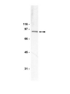
Western Blotting Analysis: 1.0 µg/mL of this antibody detected the full-length and cleaved forms of PTPRT in rat brain tissue lysate.
Western Blotting Analysis: 0.5 µg/mL of this antibody detected the full-length and a cleaved form of PTPRT in brain and liver homogenates from wild-type, but not PTPRT-knockout mice (Courtesy of Dr. Zhenghe Wang, Case Western Reserve University, Cleveland, OH).
Western Blotting Analysis: 2.0 µg/mL of this antibody detected the full-length and a cleaved form of PTPRT in small intestine epithelium homogenate from wild-type mice (Courtesy of Dr. Zhenghe Wang, Case Western Reserve University, Cleveland, OH).
Western Blotting Analysis: A representative lot detected exogenously expressed human PTPRT using transiently transfected HNCC CAL-33 (squamous carcinomas of the tongue) cells expressing wild-type, PTPase domain mutatant (A1022E), or FN3-domain mutatant (P497T) PTPRT, as well as in transfected HNSCC PCI-52-SD1 cells stably expressing wild-type human PTPRT (Lui, V.W., et al. (2014). Proc. Natl. Acad. Sci. U.S.A. 111(3):1114-1119).

Qualità

Evaluated by Western Blotting in mouse brain tissue lysate.

Western Blotting Analysis: 0.5 µg/mL of this antibody detected the full-length and a cleaved form of PTPRT in mouse brain tissue lysate.

Descrizione del bersaglio

~ 190/90 kDa observed. Due to glycosylation, the full-length target band (~190 kDa) appears larger than the calculated molecular weight (162.1 kDa). Cleaved/truncated forms between ~70 kDa to 120 kDa can also be detected. It is believed that PTPRT is proteolytically cleaved upon activation. The intracellular portion containing the two protein tyrosine phosphatase (PTPase) domains then translocates into nucleus, where it dephosphorylates STAT3 (Zhang, X., et al. (2007). Proc. Natl. Acad. Sci. U.S.A. 104(10):4060-4064).

Stato fisico

Format: Purified
Protein G Purified
Purified mouse monoclonal IgG1κ antibody in buffer containing 0.1 M Tris-Glycine (pH 7.4), 150 mM NaCl with 0.05% sodium azide.

Stoccaggio e stabilità

Stable for 1 year at 2-8°C from date of receipt.

Altre note

Concentration: Please refer to lot specific datasheet.

Esclusione di responsabilità

Unless otherwise stated in our catalog or other company documentation accompanying the product(s), our products are intended for research use only and are not to be used for any other purpose, which includes but is not limited to, unauthorized commercial uses, in vitro diagnostic uses, ex vivo or in vivo therapeutic uses or any type of consumption or application to humans or animals.

Not finding the right product?  

Try our Motore di ricerca dei prodotti.

Codice della classe di stoccaggio

12 - Non Combustible Liquids

Classe di pericolosità dell'acqua (WGK)

WGK 1

Punto d’infiammabilità (°F)

Not applicable

Punto d’infiammabilità (°C)

Not applicable


Certificati d'analisi (COA)

Cerca il Certificati d'analisi (COA) digitando il numero di lotto/batch corrispondente. I numeri di lotto o di batch sono stampati sull'etichetta dei prodotti dopo la parola ‘Lotto’ o ‘Batch’.

Possiedi già questo prodotto?

I documenti relativi ai prodotti acquistati recentemente sono disponibili nell’Archivio dei documenti.

Visita l’Archivio dei documenti

Frequent mutation of receptor protein tyrosine phosphatases provides a mechanism for STAT3 hyperactivation in head and neck cancer.
Lui, VW; Peyser, ND; Ng, PK; Hritz, J; Zeng, Y; Lu, Y; Li, H; Wang, L; Gilbert et al.
Proceedings of the National Academy of Sciences of the USA null

Il team dei nostri ricercatori vanta grande esperienza in tutte le aree della ricerca quali Life Science, scienza dei materiali, sintesi chimica, cromatografia, discipline analitiche, ecc..

Contatta l'Assistenza Tecnica.