MABN878
Anti-phospho-Amyloid beta (Ser8) Antibody, clone 1E4E11
clone 1E4E11, from mouse
Sinonimo/i:
Aβ peptide, Ser8 phosphorylated, Abeta peptide, Ser8 phosphorylated, Amyloid β peptide, Ser8 phosphorylated, Aβ1-40, Ser8 phosphorylated, Aβ1-42, Ser8 phosphorylated, Abeta1-40, Ser8 phosphorylated, Abeta1-42, Ser8 phosphorylated
About This Item
Prodotti consigliati
Origine biologica
mouse
Livello qualitativo
Forma dell’anticorpo
purified immunoglobulin
Tipo di anticorpo
primary antibodies
Clone
1E4E11, monoclonal
Reattività contro le specie
human
tecniche
ELISA: suitable
immunofluorescence: suitable
immunohistochemistry: suitable
western blot: suitable
Isotipo
IgG1κ
N° accesso NCBI
N° accesso UniProt
modifica post-traduzionali bersaglio
phosphorylation (pSer8)
Informazioni sul gene
human ... APP(351)
Descrizione generale
Specificità
Immunogeno
Applicazioni
Neuroscience
Neurodegenerative Diseases
Western Blotting Analysis: 2 µg/mL from a representative lot detected phosphorylated oligomeric amyloid beta (pAβ) peptides in 50 µg of brain extract from an 8-month old APP/PS1KI transgenic mouse, but not in extract from an age-matched non-transgenic mouse (Courtesy of Dr. Kumar and Prof. Dr. Walter, Department of Neurology, University Bonn, Germany).
ELISA Analysis: Clone 1E4E11 hybridoma culture supernatant detected the immunogen peptide with phosphorylated Ser8, but not the corresponding non-phosphorylated peptide (Kumar, S., et al. (2013). Acta Neuropathol. 125(5):699-709).
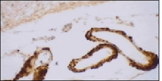
Immunohistochemistry Analysis: A representative lot detected age-dependent phospho-amyloid beta (pAβ) immunoreactivity and localization in paraffin-embedded APP/PS1KI mouse brain sections following antigen retrieval by heat and 88% formic acid treatments (Kumar, S., et al. (2013). Acta Neuropathol. 125(5):699-709).
Immunofluorescence Analysis: A representative lot detected phospho-amyloid beta (pAβ) immunoreactivity in cortical neurons colocalized with that detected with a non-phospho-specific Aβ antibody by dual fluorescent immunohistochemistry staining of paraffin-embedded 2-month old APP/PS1KI mice cotex sections following anigen retrieval by heat and 88% formic acid treatments (Kumar, S., et al. (2013). Acta Neuropathol. 125(5):699-709).
Western Blotting Analysis: A representative lot detected monomeric, dimeric, trimeric, and oligomeric forms of synthetic Aβ1-40 and Aβ1-42 peptides with phosphorylated Ser8, but not the non-phosphorylated Aβ1-40 and Aβ1-42 peptides (Kumar, S., et al. (2013). Acta Neuropathol. 125(5):699-709).
Western Blotting Analysis: A representative lot detected monomeric, dimeric, and oligomeric forms of phosphorylated amyloid beta (pAβ) peptides in brain extracts from 6- and 12-month old APP/PS1KI transgenic mice, but not in brain extracts from age-matched non-transgenic mice (Kumar, S., et al. (2013). Acta Neuropathol. 125(5):699-709).
Note: Formic acid (88%) treatment following heat retrieval is recommended for immunohistochemical detection of aggregated intraneuronal Abeta peptides in brain sections (Kumar, S., et al. (2013). Acta Neuropathol. 125(5):699-709; Christensen, D.Z., et al. (2009). Brain Res. 1301:116-125).
Qualità
Isotyping Analysis: The identity of this monoclonal antibody is confirmed by isotyping test to be IgG1κ.
Descrizione del bersaglio
Stato fisico
Stoccaggio e stabilità
Altre note
Esclusione di responsabilità
Non trovi il prodotto giusto?
Prova il nostro Motore di ricerca dei prodotti.
Codice della classe di stoccaggio
12 - Non Combustible Liquids
Classe di pericolosità dell'acqua (WGK)
WGK 1
Punto d’infiammabilità (°F)
Not applicable
Punto d’infiammabilità (°C)
Not applicable
Certificati d'analisi (COA)
Cerca il Certificati d'analisi (COA) digitando il numero di lotto/batch corrispondente. I numeri di lotto o di batch sono stampati sull'etichetta dei prodotti dopo la parola ‘Lotto’ o ‘Batch’.
Possiedi già questo prodotto?
I documenti relativi ai prodotti acquistati recentemente sono disponibili nell’Archivio dei documenti.
Il team dei nostri ricercatori vanta grande esperienza in tutte le aree della ricerca quali Life Science, scienza dei materiali, sintesi chimica, cromatografia, discipline analitiche, ecc..
Contatta l'Assistenza Tecnica.