
MABF245
Anti-TIM-1 Antibody, clone 3B3
clone 3B3, from rat
Sinonimo/i:
Hepatitis A virus cellular receptor 1 homolog, HAVcr-1, Kidney injury molecule 1, KIM-1, T cell immunoglobulin and mucin domain-containing protein 1, T-cell immunoglobulin mucin receptor 1, T cell membrane protein 1, TIM-1, TIMD-1
About This Item
Prodotti consigliati

Origine biologica
rat
Livello qualitativo
Forma dell’anticorpo
purified antibody
Tipo di anticorpo
primary antibodies
Clone
3B3, monoclonal
Reattività contro le specie
mouse
tecniche
flow cytometry: suitable
Isotipo
IgG2aκ
N° accesso NCBI
N° accesso UniProt
Condizioni di spedizione
ambient
modifica post-traduzionali bersaglio
unmodified
Informazioni sul gene
human ... HAVCR1(26762)
Descrizione generale
Specificità
Immunogeno
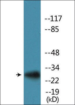
Applicazioni
Affects Function: A representative lot induced a rapid capping of anti-CD3 bound CD3 on the surface of CD4+ T cells with a concomitant T cell morphology change and increased motility (Xiao, S., et al. (2007). J. Exp. Med. 204(7):1691-1702).
Affects Function: A representative lot enhanced the severity of experimental autoimmune encephalomyelitis (EAE) in mice, while clone RMT1-10 prevented EAE (Xiao, S., et al. (2007). J. Exp. Med. 204(7):1691-1702).
Affects Function: A representative lot enhanced T cell proliferation by crosslinking TIM-1 in vitro. When injected in mice in vivo at the beginning and during OVA sensitization, clone 3B3 prevented tolerance induction and enhanced response of isolated spleen T cells to OVA stimulation in culture (Umetsu, S.E., et al. (2005). Nat. Immunol.6(5):447-454).
Affinity Binding Assay: A representative lot bound TIM-1 with a similar association rate as clone RMT1-10 (Cat. No. MABF225), while clone 3B3 displayed a 10-times slower dissociation rate than clone RMT1-10 (Xiao, S., et al. (2007). J. Exp. Med. 204(7):1691-1702).
ELISA Analysis: Representative lots detected recombinant murine TIM-1, but not TIM-3 or TIM-4, Ig fusion protein. Clone 3B3 displayed an enhanced affinity toward TIM-4 fusion construct lacking the mucin domain when compared to full-length TIM-4 fusion construct (Xiao, S., et al. (2007). J. Exp. Med. 204(7):1691-1702; Umetsu, S.E., et al. (2005). Nat. Immunol.6(5):447-454).
Flow Cytometry Analysis: A representative lot immunostained the surface of TIM-1-transfected, but not untransfected or TIM-2-transfected 300.19 mouse pre-B-cells. A time-dependent increase of CD4+ cell surface TIM-1 expression was detected following TCR activation by APCs or by anti-CD3 and anti-CD28 antibodies (Umetsu, S.E., et al. (2005). Nat. Immunol.6(5):447-454).
Inflammation & Immunology
Qualità
Flow Cytometry Analysis: 0.5 µg of this antibody detected the exogenously expressed murine TIM-1 on the surface of one million transfected 300.19 mouse pre-B-cells.
Descrizione del bersaglio
Stato fisico
Stoccaggio e stabilità
Handling Recommendations: Upon receipt and prior to removing the cap, centrifuge the vial and gently mix the solution. Aliquot into microcentrifuge tubes and store at -20°C. Avoid repeated freeze/thaw cycles, which may damage IgG and affect product performance.
Altre note
Esclusione di responsabilità
Non trovi il prodotto giusto?
Prova il nostro Motore di ricerca dei prodotti.
Codice della classe di stoccaggio
12 - Non Combustible Liquids
Classe di pericolosità dell'acqua (WGK)
WGK 2
Punto d’infiammabilità (°F)
Not applicable
Punto d’infiammabilità (°C)
Not applicable
Certificati d'analisi (COA)
Cerca il Certificati d'analisi (COA) digitando il numero di lotto/batch corrispondente. I numeri di lotto o di batch sono stampati sull'etichetta dei prodotti dopo la parola ‘Lotto’ o ‘Batch’.
Possiedi già questo prodotto?
I documenti relativi ai prodotti acquistati recentemente sono disponibili nell’Archivio dei documenti.
Il team dei nostri ricercatori vanta grande esperienza in tutte le aree della ricerca quali Life Science, scienza dei materiali, sintesi chimica, cromatografia, discipline analitiche, ecc..
Contatta l'Assistenza Tecnica.