Passa al contenuto
Merck
Tutte le immagini(1)

Documenti fondamentali

MAB3747-I

Sigma-Aldrich

Anti-Microphthalmia (Mi) Antibody, clone C5

clone C5, from mouse

Sinonimo/i:

Microphthalmia-associated transcription factor, Class E basic helix-loop-helix protein 32, bHLHe32

Autenticatiper visualizzare i prezzi riservati alla tua organizzazione & contrattuali


About This Item

Codice UNSPSC:
12352203
eCl@ss:
32160702
NACRES:
NA.41

Origine biologica

mouse

Livello qualitativo

Forma dell’anticorpo

purified immunoglobulin

Tipo di anticorpo

primary antibodies

Clone

C5, monoclonal

Reattività contro le specie

mouse, human

Reattività contro le specie (prevista in base all’omologia)

rat

tecniche

electrophoretic mobility shift assay: suitable
immunocytochemistry: suitable
immunofluorescence: suitable
immunohistochemistry: suitable
immunoprecipitation (IP): suitable
western blot: suitable

Isotipo

IgG2aκ

N° accesso NCBI

N° accesso UniProt

Condizioni di spedizione

wet ice

modifica post-traduzionali bersaglio

unmodified

Informazioni sul gene

human ... MITF(4286)

Descrizione generale

MiTF (Microphthalmia associated transcription factor) is a basic helix loop helix leucine zipper (b HLH ZIP) transcription factor implicated in pigmentation, mast cells and bone development. Mutations in MiTF cause auditory pigmentary syndromes, such as Waardenburg syndrome type II, type IIa and Tietz syndrome in humans. There are two known isoforms of MiTF differing by 66 amino acids at the NH2 terminus. Shorter forms are expressed in melanocytes and run as two bands at 52 kDa and 56 kDa, while the longer Mi form runs as a cluster of bands at 60-70 kDa in osteoclasts and in B16 melonoma cells (but not other melanoma cell lines), as well as mast cells and heart. MiTF plays a critical role in the differentiation of various cell types as neural crest-derived melanocytes, mast cells, osteoclasts and optic cup-derived retinal pigment epithelium. Mi is a basic helix-loop-helix-leucine zipper (b-HLH-ZIP) transcription factor implicated in pigmentation, mast cells and bone development. The mutation of Mi causes Waardenburg Syndrome type II in humans. In mice, a profound loss of pigmented cells in the skin eye and inner ear results, as well as osteopetrosis and defects in natural killer and mast cells. These melanocyte isoforms have been shown by two dimensional tryptic mapping to differ in c-Kit-induced phosphorylation. Osteopetrotic rat strain harbors a large genomic deletion encompassing the 3′ half of Mi including most of the b-HLH-ZIP region. Osteoclasts from these animals lack Mi protein in contrast to wild-type rat, mouse, and human osteoclasts.

Specificità

In Western blotting, it recognizes a doublet of 52-56 kDa, identified as serine-phosphorylated and unphosphorylated forms of melanocytic isoforms of microphthalmia (Mi). There are two known isoforms of Mi differing by 66 amino acids at the NH2 terminus. Shorter forms are expressed in melanocytes and run as two bands at 52 kDa and 56 kDa, while the longer Mi form runs as a cluster of bands at 60-70 kDa in osteoclasts and in B16 melonoma cells (but not other melanoma cell lines), as well as mast cells andheart. It reacts with both melanocytic as well as the nonmelanocytic isoforms of Mi. This Ab does not cross-react with other b-HLH-ZIP factors by DNA mobility shift assay.

Immunogeno

Recombinant N-terminal fragment of human microphthalmia protein.

Applicazioni

Immunohistochemistry Analysis: A representative lot detected microphthalmia immunoreactivity in formalin-fixed, paraffin-embedded human metastatic melanoma tissue sections by fluorescent immunohistochemistry (Feige, E., et. al. (2011). Proc. Natl .Acad. Sci. U. S. A. 108(43):E924-E933).
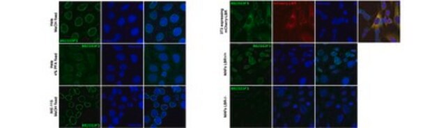
Immunocytochemistry Analysis: A representative lot detected the exogenously expressed murine microphthalmia mutant constructs, Mitf D222/236N and Mitf D222N (mi-vit), in the nucleus of transfected COS-7 cells. Dual staining showed much reduced β-catenin-anchoring ability of these mutants in the nucleus (Schepsky, A., et al. (2006). Mol. Cell. Biol. 26(23): 8914-8927).
Immunocytochemistry Analysis: A representative lot detected a time-dependent induction of microphthalmia upregulation in B16/F10 murine melanoma cells upon Forskolin stimulation by fluorescent immunocytochemistry (Bertolotto, C., et al. (1998). J. Cell Biol. 142(3):827-835).
Electrophoretic Mobility Shift Assay (EMSA): A representative lot caused a supershift of Mbox motif oligonucleotide-complexed wild-type and D222/236N and D222N mutant murine microphthalmia constructs by EMSA (Schepsky, A., et al. (2006). Mol. Cell. Biol. 26(23): 8914-8927).
Electrophoretic Mobility Shift Assay (EMSA): A representative lot caused a supershift of Mbox motif oligonucleotide-complexed microphthalmia, but not TFE3-DNA complex by EMSA using in vitro translated microphthalmia and TFE3 or B16/F10 murine melanoma cell nuclear extract (Verastegui, C., et al. (2000). Mol. Endocrinol. 14(3):449-456).
Immunoprecipitation Analysis: A representative lot immunoprecipitated microphthalmia from B16/F10 murine melanoma cell nuclear extracts (Verastegui, C., et al. (2000). Mol. Endocrinol. 14(3):449-456).
Western Blotting Analysis: A representative lot detected microphthalmia expression in murine splenocytes and B16/F10 murine melanoma cells (Verastegui, C., et al. (2000). Mol. Endocrinol. 14(3):449-456).
Western Blotting Analysis: A representative lot detected a time-dependent induction of microphthalmia upregulation in B16/F10 murine melanoma cells and normal human melanocytes upon stimulation by Forskolin or α-melanocyte–stimulating hormone (αMSH) (Bertolotto, C., et al. (1998). J. Cell Biol. 142(3):827-835).
Use Anti-Microphthalmia (Mi), clone C5 mouse monoclonal antibody validated in Electrophoretic Mobility Shift Assay (EMSA), Immunocytochemistry, Immunohistochemistry, Immunoprecipitation and Western blotting for the detection of Microphthalmia-associated transcription factor.

Qualità

Evaluated by Western Blotting in mouse brain tissue lysate.

Western Blotting Analysis: An 1:500 dilution of this antibody detected Microphthalmia in 10 µg of mouse brain tissue lysate.

Descrizione del bersaglio

~52/56 kDa observed. An uncharacterized band appears at ~140 kDa in some lysates.

Stato fisico

Format: Purified

Risultati analitici

Control
Mouse brain tissue lysates

Altre note

Concentration: Please refer to the Certificate of Analysis for the lot-specific concentration.

Non trovi il prodotto giusto?  

Prova il nostro Motore di ricerca dei prodotti.

Raccomandato

N° Catalogo
Descrizione
Determinazione del prezzo

Codice della classe di stoccaggio

12 - Non Combustible Liquids

Classe di pericolosità dell'acqua (WGK)

WGK 1

Punto d’infiammabilità (°F)

Not applicable

Punto d’infiammabilità (°C)

Not applicable


Certificati d'analisi (COA)

Cerca il Certificati d'analisi (COA) digitando il numero di lotto/batch corrispondente. I numeri di lotto o di batch sono stampati sull'etichetta dei prodotti dopo la parola ‘Lotto’ o ‘Batch’.

Possiedi già questo prodotto?

I documenti relativi ai prodotti acquistati recentemente sono disponibili nell’Archivio dei documenti.

Visita l’Archivio dei documenti

Malignant PEComa of the adrenal gland.
Lau, Sean K
Pathology, research and practice, 208, 113-117 (2012)
Ssu-Yi Lu et al.
Experimental cell research, 328(1), 32-43 (2014-08-26)
Transcription factors Mitf and NFATc1 share many downstream targets that are critical for osteoclastogenesis. Since RANKL signals induce/activate both NFATc1 and Mitf isoform-E (Mitf-E), a tissue-restricted Mitf isoform in osteoclasts, it is plausible that the two factors work together to
Hyo Jung Kim et al.
International journal of molecular sciences, 16(4), 8772-8788 (2015-04-24)
The melanin-inducing properties of cirsimaritin were investigated in murine B16F10 cells. Cirsimaritin is an active flavone with methoxy groups, which is isolated from the branches of Lithocarpus dealbatus. Tyrosinase activity and melanin content in murine B16F10 melanoma cells were increased

Il team dei nostri ricercatori vanta grande esperienza in tutte le aree della ricerca quali Life Science, scienza dei materiali, sintesi chimica, cromatografia, discipline analitiche, ecc..

Contatta l'Assistenza Tecnica.