Skip to Content
MilliporeSigma
All Photos(3)

Key Documents

483R-1

Sigma-Aldrich

p40 (ZR8) Rabbit Monoclonal Antibody

Sign Into View Organizational & Contract Pricing


About This Item

UNSPSC Code:
12352203
clone:
ZR8
application:
IHC (p)
species reactivity:
human
technique(s):
immunohistochemistry (formalin-fixed, paraffin-embedded sections): suitable (1:100-1:200 (concentrated))
citations:
8

biological source

rabbit

Quality Level

100
500

conjugate

unconjugated

antibody form

culture supernatant

antibody product type

primary antibodies

clone

ZR8

description

For In Vitro Diagnostic Use in Select Regions

form

buffered aqueous solution

species reactivity

human

packaging

vial of 0.1 mL concentrate (483R-14)
vial of 0.1 mL concentrate, Research Use Only (483R-14-RUO)
vial of 0.5 mL concentrate (483R-15)
vial of 1.0 mL concentrate (483R-16)
vial of 1.0 mL concentrate, Research Use Only (483R-16-RUO)
vial of 1.0 mL pre-dilute, Research Use Only (483R-17-RUO)
vial of 1.0 mL pre-dilute, ready to use (483R-17)
vial of 25.0 mL pre-dilute, ready-to-use (483R-10)
vial of 25.0 mL pre-dilute, ready-to-use, Research Use Only (483R-10-RUO)
vial of 7.0 mL pre-dilute, ready to use (483R-18)
vial of 7.0 mL pre-dilute, ready to use, Research Use Only (483R-18-RUO)

manufacturer/tradename

Cell Marque

technique(s)

immunohistochemistry (formalin-fixed, paraffin-embedded sections): suitable (1:100-1:200 (concentrated))

isotype

IgG

control

lung (squamous cell carcinoma), prostate, urothelial carcinoma

shipped in

wet ice

storage temp.

2-8°C

visualization

nuclear

Related Categories

General description

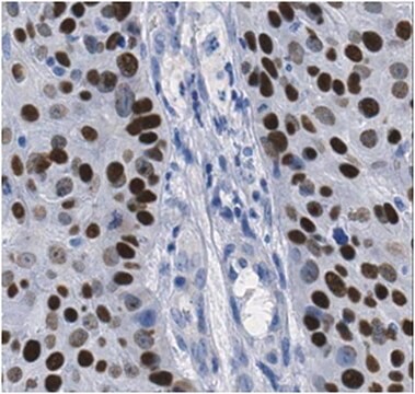
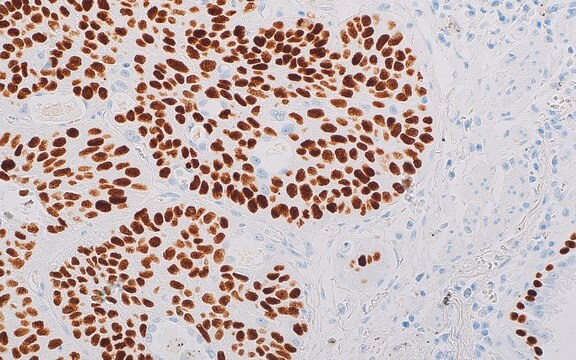
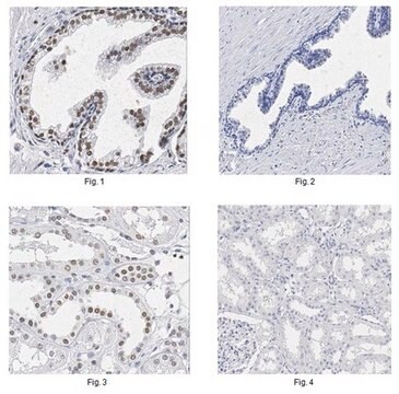
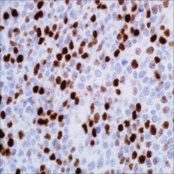
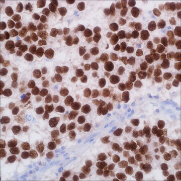
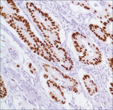
p40 is an isoform of p63, a transcription factor that regulates many cell activities, including cell proliferation, maintenance, and differentiation. It performs as a sensitive and specific tool for aiding in the identification of squamous cell carcinoma of the lung. In addition to its utility as a squamous differentiation marker, p40 has also been proven to be a valuable marker for highlighting myoepithelial and basal cell populations in prostate, breast, skin, and salivary gland.1-5 Strong p40 expression is frequently observed in esophageal cancerous squamous lesions.6 Immunohistochemical detection of p40 can also be helpful in identifying urothelial carcinoma.7 In cases of prostate carcinoma, p40 is almost always found to be negative for basal cell staining.8

Quality

  • United States - IVD
  • Canada - RUO
  • European Union - IVD
  • Japan - RUO

Physical form

Solution in Tris Buffer, pH 7.3-7.7, with 1% BSA and <0.1% Sodium Azide.

Preparation Note

Download the IFU specific to your product lot and format
Note: This requires a keycode which can be found on your packaging or product label.

Other Notes

For Technical Service please contact: 800-665-7284 or email: service@cellmarque.com

Not finding the right product?  

Try our Product Selector Tool.

Storage Class Code

12 - Non Combustible Liquids

WGK

WGK 2

Flash Point(F)

Not applicable

Flash Point(C)

Not applicable


Choose from one of the most recent versions:

Certificates of Analysis (COA)

Lot/Batch Number

Sorry, we don't have COAs for this product available online at this time.

If you need assistance, please contact Customer Support.

Already Own This Product?

Find documentation for the products that you have recently purchased in the Document Library.

Visit the Document Library

Hermann Brustmann
Pathology research international, 2015, 897927-897927 (2015-04-09)
Immunohistochemistry is important for the accurate diagnosis of basal cells in atypical glandular proliferations of the prostate. p40, an isoform of p63, may be an adjunct to a marker panel in this setting. Biopsies of 68 patients were analyzed by
Khairunisa Ahmad Affandi et al.
Journal of pathology and translational medicine, 52(5), 283-289 (2018-09-22)
Lung cancer is the third most common cancer worldwide. With major advances in the molecular testing of lung cancers and the introduction of targeted therapies, the distinction between adenocarcinoma and squamous cell carcinoma as well as pathologic subtyping has become
Helene Geddert et al.
Human pathology, 34(9), 850-856 (2003-10-17)
p63, a member of the p53 gene family, is known to encode functionally antagonistic protein isoforms. Although transactivating protein isoforms display p53-like functions, deltaNp63 isoforms act toward p53 in a dominant negative way. Using immunohistochemistry, we examined the expression of
Ritika Walia et al.
The Indian journal of medical research, 146(1), 42-48 (2017-11-24)
Accurate histopathological subtyping of non-small cell lung carcinoma (NSCLC) is essential for targeted therapeutic agents. Immunohistochemistry (IHC) is helpful in identification of different tumour subtypes. In this study two marker approaches, one each for glandular and squamous cell differentiation was

Our team of scientists has experience in all areas of research including Life Science, Material Science, Chemical Synthesis, Chromatography, Analytical and many others.

Contact Technical Service