MABT397
Anti-CEACAM1/CD66a Antibody, clone B3-17
clone B3-17, from mouse
Sinônimo(s):
Carcinoembryonic antigen-related cell adhesion molecule 1, Biliary glycoprotein 1, BGP-1, CD66a
About This Item
Produtos recomendados
fonte biológica
mouse
Nível de qualidade
forma do anticorpo
purified immunoglobulin
tipo de produto de anticorpo
primary antibodies
clone
B3-17, monoclonal
reatividade de espécies
human
técnica(s)
ELISA: suitable
flow cytometry: suitable
immunocytochemistry: suitable
immunohistochemistry: suitable (paraffin)
western blot: suitable
Isotipo
IgG1κ
nº de adesão NCBI
nº de adesão UniProt
Condições de expedição
wet ice
modificação pós-traducional do alvo
unmodified
Informações sobre genes
human ... CEACAM1(634)
Descrição geral
Imunogênio
Aplicação
Flow Cytometry Analysis: 10 µg/mL from a representative lot detected the exogenously expressed human CEACAM1/CD66a on the surface of transfected HeLa cells (Courtesy of Dr. B. Singer, University Duisburg-Essen, Germany).
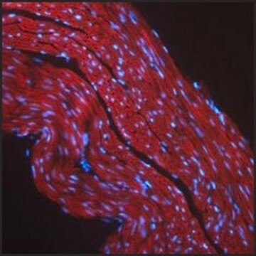
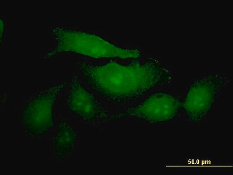
Immunocytochemistry Analysis: 10 µg/mL from a representative lot detected CEACAM1/CD66a on the surface of HT-29 human colorectal carcinoma cells (Courtesy of Dr. B. Singer, University Duisburg-Essen, Germany).
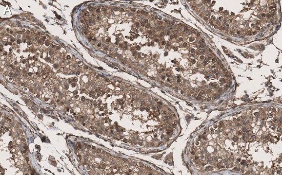
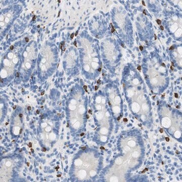
Immunohistochemistry Analysis: 10 µg/mL from a representative lot detected CEACAM1/CD66a immunoreactivity in human jejunum tissue (Courtesy of Dr. B. Singer, University Duisburg-Essen, Germany).
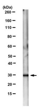
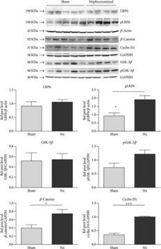
Western Blotting Analysis: 10 µg/mL from a representative lot detected the exogensously expressed human CEACAM1/CD66a extracellular domain-Fc fusion in lysates from transfected cells (Courtesy of Dr. B. Singer, University Duisburg-Essen, Germany).
Flow Cytometry Analysis: Representative lots, either unconjugated or FITC-conjugated, detected CECAM1 immunoreactivity on the surface of human peripheral blood naïve and memory B-cells (Khairnar, V., et al. (2015). Nat. Commun. 6:6217; Seifert, M., et al. (2015). Proc. Natl. Acad. Sci. 112(6):E546-555).
Flow Cytometry Analysis: A representative lot detected CEACAM1/CD66a induction on the surface of normal human bronchial epithelial (NHBE) cells stimulated with poly(I:C) or interferons (Klaile, E., et al. (2013) Respir Res. 14:85).
Flow Cytometry Analysis: A representative lot detected CEACAM1/CD66a expression on the surface of 2 day-starved human epithelial (HT29, T102/3) and endothelial (AS-M.5) cells, as well as multivesicular bodies (MVBs) derived from these cells (Muturi H.T., et al. (2013) PLoS One. 8(9):e74654).
Immunocytochemistry Analysis: A representative lot detected CEACAM1/CD66a immunoreactivity on the surface of normal human bronchial epithelial (NHBE) cells by indirect immunofluorescence staining (Klaile, E., et al. (2013) Respir Res. 14:85).
Immunohistochemistry Analysis: A representative lot detected CEACAM1/CD66a in paraffin-embedded human lung cancer tissue sections (Klaile, E., et al. (2013) Respir Res. 14:85).
Western Blot Analysis: A representative lot detected CEACAM1/CD66a in 2 day-starved AS-M.5 human endothelialn cells and AS-M.5-derived multivesicular bodies (MVBs) (Muturi H.T., et al. (2013) PLoS One. 8(9):e74654).
Qualidade
Western Blotting Analysis: 2 µg/mL of this antibody detected CEACAM1/CD66a in 10 µg of HepG2 cell lysate.
Descrição-alvo
forma física
Outras notas
Não está encontrando o produto certo?
Experimente o nosso Ferramenta de seleção de produtos.
Código de classe de armazenamento
12 - Non Combustible Liquids
Classe de risco de água (WGK)
WGK 1
Ponto de fulgor (°F)
Not applicable
Ponto de fulgor (°C)
Not applicable
Certificados de análise (COA)
Busque Certificados de análise (COA) digitando o Número do Lote do produto. Os números de lote e remessa podem ser encontrados no rótulo de um produto após a palavra “Lot” ou “Batch”.
Já possui este produto?
Encontre a documentação dos produtos que você adquiriu recentemente na biblioteca de documentos.
Nossa equipe de cientistas tem experiência em todas as áreas de pesquisa, incluindo Life Sciences, ciência de materiais, síntese química, cromatografia, química analítica e muitas outras.
Entre em contato com a assistência técnica