Pular para o conteúdo
Merck
Todas as fotos(1)

Documentos Principais

MAB16985F

Sigma-Aldrich

Anti-MCAM Antibody, clone P1H12, FITC conjugated

clone P1H12, Chemicon®, from mouse

Sinônimo(s):

MUC18, CD146

Faça loginpara ver os preços organizacionais e de contrato


About This Item

Código UNSPSC:
12352203
eCl@ss:
32160702
NACRES:
NA.41

fonte biológica

mouse

Nível de qualidade

conjugado

FITC conjugate

forma do anticorpo

purified immunoglobulin

tipo de produto de anticorpo

primary antibodies

clone

P1H12, monoclonal

reatividade de espécies

canine, mouse, human

não deve reagir com

rat

fabricante/nome comercial

Chemicon®

técnica(s)

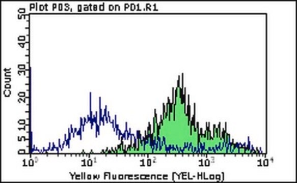
flow cytometry: suitable

Isotipo

IgG1

nº de adesão NCBI

nº de adesão UniProt

Condições de expedição

wet ice

modificação pós-traducional do alvo

unmodified

Informações sobre genes

human ... MCAM(4162)

Descrição geral

MUC18 antigen (CD146), a member of the immuoglobulin superfamily, is an intrinsic membrane glycoprotein of 110-120 kDa found on the surface of endothelial cells, bone marrow fibroblasts and various melanomas. MUC18 has been used as a marker of tumor progression in human melanoma because expression in those tumors correlates strongly with poor prognosis and the development of metastic disease. In addition, a number of human T, B and myeloid leukemic cell lines seem to express MUC18. The close structural relationship with N-CAM and related molecules suggests that MUC18 may be also a developmentally regulated cell adhesion molecule (Melanoma adhesion molecule or MCAM).

Especificidade

Detects circulating endothelial cells in human. Expected to cross-react with dog & mouse.
P1H12 reacts specifically with CD146 {MCAM, MUC18}. In blood and marrow P1H12 reacts only with endothelial cells, and has been used to detect circulating endothelial cells in human, dogs and mouse samples. It positively stains normal, primary HUVEC and MVEC cultures and the endothelial cells of all vessels in normal frozen sections of human skin, intestine, ovary tonsil, lymph node, lung, and kidney. It does not stain carcinoma cell lines HT-29 and COLO205, and M21, the T cell lines Jurkat and HuT78, fibroblasts, HL-60 or CHO cells, or EBV-transformed B cell lines, although it expected that CD146 expression is present in many tumor lines. P1H12 does not stain normal monocytes, granulocytes, red cells, platelets, T cells or B cells from marrow or peripheral blood; nor does it detect marrow megakaryocytes or the megakaryoblast line HU3. The peripheral blood cells that do stain with P1H12 also are positive for both von Willebrand Factor (vWF) and thrombomodulin (the combined expression of which is limited to endothelium), and they stain for flt and flk. In recent testing, melanoma A2058, SKMEL.3 and A375.S2 are positive, while murine melanoma M3 is negative. One smooth muscle line (HISM) was positive and one negative (TIGHA-VSMC). Human blood lymphoctes are negative with or without prior stimulation.

Imunogênio

Immunization of mice with HUVEC

Aplicação

Detect MCAM using this Anti-MCAM Antibody, clone P1H12, FITC conjugated validated for use in FC.
Immunocytochemistry: 1-10 μg/mL. Works best on EDTA or Trypsin lifted endothelial cells.

Immunohistochemistry: 1-10 μg/mL. 4% PFA for 30min RT or <2hrs @4°C. Block w/ 1%BSA/0.2%tween20/PBC for 30min. Works well in frozen tissue; fixed or unfixed.

Facs Analysis: 1-10 μg/mL

Optimal working dilutions and protocols must be determined by end user.
Research Category
Cell Structure
Research Sub Category
Adhesion (CAMs)

forma física

Protein A purified
Purified immunoglobulin. Liquid in 0.02M PBS, 0.25M NaCl, pH=7.6, 15 mg/nL BSA containing 0.1% sodium azide as a preservative.

Armazenamento e estabilidade

Maintain for 1 year at 2–8°C from date of shipment. For maximum recovery of product, centrifuge the original vial after thawing and prior to removing the cap.

Nota de análise

Control
HUVEC cells

Outras notas

Concentration: Please refer to the Certificate of Analysis for the lot-specific concentration.

Informações legais

CHEMICON is a registered trademark of Merck KGaA, Darmstadt, Germany

Exoneração de responsabilidade

Unless otherwise stated in our catalog or other company documentation accompanying the product(s), our products are intended for research use only and are not to be used for any other purpose, which includes but is not limited to, unauthorized commercial uses, in vitro diagnostic uses, ex vivo or in vivo therapeutic uses or any type of consumption or application to humans or animals.

Não está encontrando o produto certo?  

Experimente o nosso Ferramenta de seleção de produtos.

Código de classe de armazenamento

12 - Non Combustible Liquids

Classe de risco de água (WGK)

WGK 2

Ponto de fulgor (°F)

Not applicable

Ponto de fulgor (°C)

Not applicable


Certificados de análise (COA)

Busque Certificados de análise (COA) digitando o Número do Lote do produto. Os números de lote e remessa podem ser encontrados no rótulo de um produto após a palavra “Lot” ou “Batch”.

Já possui este produto?

Encontre a documentação dos produtos que você adquiriu recentemente na biblioteca de documentos.

Visite a Biblioteca de Documentos

Phase II study of Cediranib (AZD 2171), an inhibitor of the vascular endothelial growth factor receptor, for second-line therapy of small cell lung cancer (National Cancer Institute #7097).
Ramalingam, SS; Belani, CP; Mack, PC; Vokes, EE; Longmate, J; Govindan, R; Koczywas et al.
Journal of Thoracic Oncology : Official Publication of the International Association for the Study of Lung Cancer null
The Schneiderian membrane contains osteoprogenitor cells: in vivo and in vitro study.
S Srouji, T Kizhner, D Ben David, M Riminucci, P Bianco, E Livne
Calcified Tissue International null
Stefania Elena Navone et al.
Vascular cell, 5(1), 10-10 (2013-05-16)
Brain microvascular endothelial cells (BMVECs) constitute the primary limitation for passage of ions and molecules from the blood into the brain through the blood brain barrier. Numerous multi-step procedures for isolating and culturing BMVECs have been described. However, each one
PLXDC1 (TEM7) is identified in a genome-wide expression screen of glioblastoma endothelium.
Robert M Beaty,Jennifer B Edwards,Kathy Boon,I-Mei Siu,James E Conway,Gregory J Riggins
Journal of Neuro-Oncology null
Nadia Quirici et al.
Stem cells and development, 19(6), 915-925 (2009-11-26)
Stem cells hold great promise in tissue engineering for repairing tissues damaged by disease or injury. Mesenchymal stem cells (MSCs) are multipotent cells able to proliferate and differentiate into multiple mesodermal tissues such as bone, cartilage, muscle, tendon, and fat.

Nossa equipe de cientistas tem experiência em todas as áreas de pesquisa, incluindo Life Sciences, ciência de materiais, síntese química, cromatografia, química analítica e muitas outras.

Entre em contato com a assistência técnica