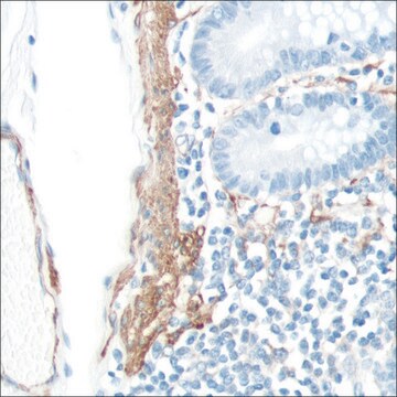
393A-1
Adipophilin Rabbit Polyclonal Antibody
About This Item
Produits recommandés
Source biologique
rabbit
Niveau de qualité
100
500
Conjugué
unconjugated
Forme d'anticorps
Ig fraction of antiserum
Type de produit anticorps
primary antibodies
Clone
polyclonal
Description
For In Vitro Diagnostic Use in Select Regions (See Chart)
Forme
buffered aqueous solution
Espèces réactives
human
Conditionnement
vial of 0.1 mL concentrate (393A-14)
vial of 0.5 mL concentrate (393A-15)
bottle of 1.0 mL predilute (393A-17)
vial of 1.0 mL concentrate (393A-16)
bottle of 7.0 mL predilute (393A-18)
Fabricant/nom de marque
Cell Marque™
Technique(s)
immunohistochemistry (formalin-fixed, paraffin-embedded sections): 1:25-1:100
Contrôle
sebaceous neoplasms (intracytoplasmic lipid droplet)
Conditions d'expédition
wet ice
Température de stockage
2-8°C
Visualisation
membranous
Informations sur le gène
human ... PLIN2(123)
Description générale
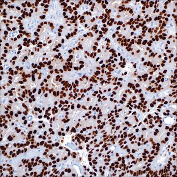
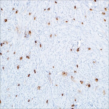
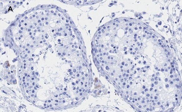
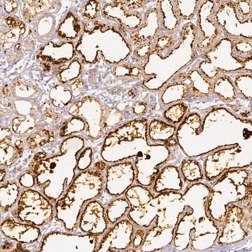
Of 25 sebaceous carcinomas, 23 (92%) were also labeled with a similar pattern. Additionally, in cases of poorly differentiated sebaceous carcinoma (n=11), in which sebaceous differentiation could not have been reliably interpreted in H&E sections, adipophilin highlighted the sebocytes with a strong membranous labeling of intracytoplasmic lipid droplets in 9 of 11 cases (82%). Moreover, 10 of 10 (100%) xanthelasmas, 9 of 10 (90%) xanthogranulomas, 6 of 6 (100%) xanthomas, and 9 of 13 (63%) metastatic renal cell carcinomas were also weakly-to- moderately positive for adipophilin. Expression of adipophilin with a membranous pattern of staining was not seen in any of the other clear cell lesions of the skin, including basal and squamous cell carcinomas, trichilemmomas, clear cell hidradenomas, or balloon cell nevi. Interestingly, a nonspecific granular uptake of anti-adipophilin was seen in adjacent macrophages, keratohyalin granules of epithelial squamous cells, and some tumor cells. Therefore, this anti-adipophilin is suitable for immunostaining formalin-fixed, paraffin-embedded tissue and is helpful in the identification of intracytoplasmic lipids, as seen in sebaceous lesions. It is especially helpful in identifying intracytoplasmic lipid vesicles in poorly differentiated sebaceous carcinomas in challenging cases such as small periocular biopsy specimens.
Qualité
 IVD |  IVD |  IVD |  RUO |
Liaison
Forme physique
Notes préparatoires
Autres remarques
Informations légales
Vous ne trouvez pas le bon produit ?
Essayez notre Outil de sélection de produits.
Certificats d'analyse (COA)
Recherchez un Certificats d'analyse (COA) en saisissant le numéro de lot du produit. Les numéros de lot figurent sur l'étiquette du produit après les mots "Lot" ou "Batch".
Déjà en possession de ce produit ?
Retrouvez la documentation relative aux produits que vous avez récemment achetés dans la Bibliothèque de documents.
Articles
Immunohistochemistry (IHC) techniques and applications have greatly improved, dermatopathology is still largely based on H&E stained slides.This paper outlines ways in which IHC antibodies can be utilized for dermatopathology.
Notre équipe de scientifiques dispose d'une expérience dans tous les secteurs de la recherche, notamment en sciences de la vie, science des matériaux, synthèse chimique, chromatographie, analyse et dans de nombreux autres domaines..
Contacter notre Service technique