Accéder au contenu
Merck
Toutes les photos(1)

Key Documents

380R-2

Sigma-Aldrich

Arginase-1 (EP261) Rabbit Monoclonal Primary Antibody

Se connecterpour consulter vos tarifs contractuels et ceux de votre entreprise/organisme


About This Item

Code UNSPSC :
12352203

Source biologique

rabbit

Niveau de qualité

100
500

Conjugué

unconjugated

Forme d'anticorps

culture supernatant

Type de produit anticorps

primary antibodies

Clone

EP261, monoclonal

Description

For In Vitro Diagnostic Use in Select Regions

Forme

buffered aqueous solution

Espèces réactives

human

Conditionnement

vial of 0.1 mL concentrate (380R-24)
vial of 0.1 mL concentrate Research Use Only (380R-24-RUO)
vial of 0.5 mL concentrate (380R-25)
vial of 1.0 mL concentrate (380R-26)
vial of 1.0 mL concentrate Research Use Only (380R-26-RUO)
vial of 1.0 mL pre-dilute Research Use Only (380R-27-RUO)
vial of 1.0 mL pre-dilute ready-to-use (380R-27)
vial of 7.0 mL pre-dilute ready-to-use (380R-28)
vial of 7.0 mL pre-dilute ready-to-use Research Use Only (380R-28-RUO)

Fabricant/nom de marque

Cell Marque

Technique(s)

immunohistochemistry (formalin-fixed, paraffin-embedded sections): 1:50-1:200 (concentrated)

Isotype

IgG

Contrôle

hepatocellular carcinoma, normal liver

Conditions d'expédition

wet ice

Température de stockage

2-8°C

Visualisation

cytoplasmic, nuclear

Informations sur le gène

human ... ARG1(383)

Description générale

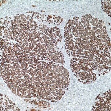
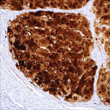
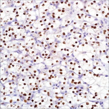
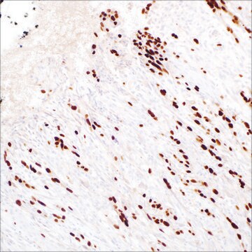
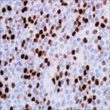
Arginase-1 is a key urea cycle metalloenzyme that has demonstrated expression in normal human liver with a high degree of specificity. Hepatocellular carcinoma (HCC) is the most common primary malignant tumor of the liver accounting for an estimated 70%-85% of total liver cancers worldwide. Diagnostic pitfalls exist in the morphologic distinction of HCC from other hepatocellular and non-hepatocellular lesions. In difficult or equivocal cases, the application of immunohistochemical (IHC) panels has been shown to aid in the distinction of benign and malignant liver lesions. In sections of normal liver, anti-arginase-1 produced strong, diffuse cytoplasmic reactivity in all hepatocytes throughout the lobule. In some cases, patchy nuclear reactivity is also evident in hepatocytes along with the cytoplasmic reactivity. Reactivity is not observed in bile duct epithelial cells, sinusoidal endothelial cells, Kupffer cells, or vascular endothelial cells. In sections of HCC, anti-arginase-1 produces cytoplasmic or cytoplasmic plus nuclear reactivity.

Qualité


IVD

IVD

IVD

RUO

Liaison

Arginase-1 Positive Control Slides, Product No. 380S, are available for immunohistochemistry (formalin-fixed, paraffin-embedded sections).

Forme physique

Solution in Tris Buffer, pH 7.3-7.7, with 1% BSA and <0.1% Sodium Azide.

Notes préparatoires

Download the IFU specific to your product lot and format
Note: This requires a keycode which can be found on your packaging or product label.

Download the latest released IFU
Note: This IFU may not apply to your specific product lot.

Autres remarques

For Technical Service please contact: 800-665-7284 or email: service@cellmarque.com

Informations légales

Cell Marque is a trademark of Merck KGaA, Darmstadt, Germany

Vous ne trouvez pas le bon produit ?  

Essayez notre Outil de sélection de produits.

Code de la classe de stockage

12 - Non Combustible Liquids

Classe de danger pour l'eau (WGK)

WGK 2

Point d'éclair (°F)

Not applicable

Point d'éclair (°C)

Not applicable


Certificats d'analyse (COA)

Recherchez un Certificats d'analyse (COA) en saisissant le numéro de lot du produit. Les numéros de lot figurent sur l'étiquette du produit après les mots "Lot" ou "Batch".

Déjà en possession de ce produit ?

Retrouvez la documentation relative aux produits que vous avez récemment achetés dans la Bibliothèque de documents.

Consulter la Bibliothèque de documents

R L Zimmerman et al.
Cancer, 93(4), 288-291 (2001-08-17)
Diagnosing liver tumors by fine-needle aspiration biopsy is safe and accurate. However, there are cases that prove diagnostically difficult. Traditionally, immunostains for alpha-fetoprotein and polyclonal carcinoembryonic antigen have been used to distinguish adenocarcinomas from hepatocellular carcinomas (HCCs). In poorly differentiated
Nehal A Radwan et al.
Diagnostic pathology, 7, 149-149 (2012-11-01)
The ability to distinguish hepatocellular carcinoma (HCC) from metastatic carcinoma (MC) involving the liver and cholangiocarcinoma (CC) by immunohistochemistry has been limited by the lack of a reliable positive marker for hepatocellular differentiation. Arginase-1 is a marker for HCC recently
T H Niemann et al.
Cancer, 87(5), 295-298 (1999-10-28)
Fine-needle aspiration biopsy (FNAB) is frequently used to diagnose mass lesions in the liver. Differentiating metastatic adenocarcinoma from primary hepatocellular carcinoma can be difficult. Despite a number of morphologic criteria, there remain occasional cases in which the cytologic features fail
Alexandre Sherlley Casimiro Onofre et al.
Cancer, 111(4), 259-268 (2007-06-15)
Difficulties with cytologic diagnoses on fine-needle aspiration cytology (FNAC) of the liver can be overcome by the application of immunocytochemical panels applied on smears. The aim of the current study was to analyze the performance of a panel of monoclonal
Ahmedin Jemal et al.
CA: a cancer journal for clinicians, 61(2), 69-90 (2011-02-08)
The global burden of cancer continues to increase largely because of the aging and growth of the world population alongside an increasing adoption of cancer-causing behaviors, particularly smoking, in economically developing countries. Based on the GLOBOCAN 2008 estimates, about 12.7

Notre équipe de scientifiques dispose d'une expérience dans tous les secteurs de la recherche, notamment en sciences de la vie, science des matériaux, synthèse chimique, chromatographie, analyse et dans de nombreux autres domaines..

Contacter notre Service technique