
MABT818
Anti-Myosin-3 (MYH3) Antibody, clone BF-G6
clone BF-G6, from mouse
Synonyme(s) :
Myosin-3, Muscle embryonic myosin heavy chain, Myosin heavy chain 3, Myosin heavy chain, fast skeletal muscle, embryonic, SMHCE
About This Item
Produits recommandés

Source biologique
mouse
Niveau de qualité
Forme d'anticorps
purified immunoglobulin
Type de produit anticorps
primary antibodies
Clone
BF-G6, monoclonal
Espèces réactives
rat, human
Réactivité de l'espèce (prédite par homologie)
bovine (based on 100% sequence homology)
Technique(s)
ELISA: suitable
immunocytochemistry: suitable
immunofluorescence: suitable
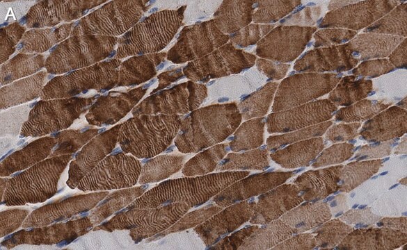
immunohistochemistry: suitable
western blot: suitable
Isotype
IgG2b, kappa
Numéro d'accès NCBI
Numéro d'accès UniProt
Conditions d'expédition
ambient
Modification post-traductionnelle de la cible
unmodified
Informations sur le gène
human ... MYH3(4621)
Description générale
Spécificité
Immunogène
Application
Cell Structure
ELISA Analysis: Clone BF-G6 hybridoma culture supernatant detected myosin from 10-week human fetus, but not myosin from 8-day new born muscle or adult skeletal muscle (Schiaffino, S., et al. (1986). Exp. Cell Res. 163(1):211-220).
Immunocytochemistry Analysis: Clone BF-G6 hybridoma culture supernatant immunostained aceton-fixed tumor cells in bone marrow aspiration from a child with rhobdomyosarcoma (Schiaffino, S., et al. (1986). Exp. Cell Res. 163(1):211-220).
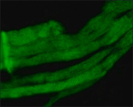
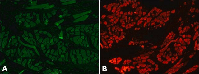
Immunofluorescence Analysis: Clone BF-G6 hybridoma culture supernatant immunostained serial transverse cryosections of rat extraocular (EO) and soleus muscles. BF-G6 staining pattern is similar, but not identical to that of MYH15 (Rossi, A.C., et al. (2010). J. Physiol. 588(Pt 2):353-364).
Immunofluorescence Analysis: Clone BF-G6 hybridoma culture supernatant immunostained frozen fetal muscle sections, while very few cells were stained in 8-week new born muscle tissue and no staining of 8-month infant muscle tissue was observed (Schiaffino, S., et al. (1986). Exp. Cell Res. 163(1):211-220).
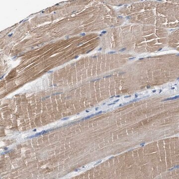
Immunohistochemistry Analysis: Clone BF-G6 hybridoma culture supernatant immunostained tumor cells in frozen human rhobdomyosarcoma sections (Schiaffino, S., et al. (1986). Exp. Cell Res. 163(1):211-220).
Western Blotting Analysis: Clone BF-G6 hybridoma culture supernatant detected myosin from 10-week human fetus, but not myosin from adult skeletal muscle (Schiaffino, S., et al. (1986). Exp. Cell Res. 163(1):211-220).
Qualité
Isotyping Analysis: The identity of this monoclonal antibody is confirmed by isotyping test to be mouse IgG2b, kappa.
Description de la cible
Forme physique
Stockage et stabilité
Autres remarques
Clause de non-responsabilité
Vous ne trouvez pas le bon produit ?
Essayez notre Outil de sélection de produits.
Code de la classe de stockage
12 - Non Combustible Liquids
Classe de danger pour l'eau (WGK)
WGK 1
Point d'éclair (°F)
Not applicable
Point d'éclair (°C)
Not applicable
Certificats d'analyse (COA)
Recherchez un Certificats d'analyse (COA) en saisissant le numéro de lot du produit. Les numéros de lot figurent sur l'étiquette du produit après les mots "Lot" ou "Batch".
Déjà en possession de ce produit ?
Retrouvez la documentation relative aux produits que vous avez récemment achetés dans la Bibliothèque de documents.
Notre équipe de scientifiques dispose d'une expérience dans tous les secteurs de la recherche, notamment en sciences de la vie, science des matériaux, synthèse chimique, chromatographie, analyse et dans de nombreux autres domaines..
Contacter notre Service technique