MAB329-C
Anti-Synaptophysin Antibody, clone SP15 (Ascites Free)
clone SP15, from mouse
Synonym(e):
Major synaptic vesicle protein p38, Synaptophysin
About This Item
Empfohlene Produkte
Biologische Quelle
mouse
Qualitätsniveau
Antikörperform
purified antibody
Antikörper-Produkttyp
primary antibodies
Klon
SP15, monoclonal
Speziesreaktivität
human, monkey, feline, rat, mouse
Methode(n)
ELISA: suitable
immunocytochemistry: suitable
immunohistochemistry: suitable
western blot: suitable
Isotyp
IgMκ
NCBI-Hinterlegungsnummer
UniProt-Hinterlegungsnummer
Posttranslationale Modifikation Target
unmodified
Angaben zum Gen
human ... SYP(6855)
Allgemeine Beschreibung
Immunogen
Anwendung
Neurowissenschaft
Entwicklungsneurowissenschaft
Western Blotting Analysis: A representative lot detected synaptophysin in rat hippocampal tissue extracts (Lin, D., et al. (2012). Behav. Brain Res. 228(2):319-327).
Western Blotting Analysis: Representative lots detected synaptophysin in mouse brain synaptosomes preparations (Shim, S.Y., et al. (2008). J. Neurosci. 28(14):3604-3614).
Western Blotting Analysis: A representative lot detected synaptophysin in the large dense-core vesicles-containing fractions obtained by sucrose gradient centrifugation of rat brain median eminence (ME) neuroterminals preparations (Yin, W., et al. (2007). Exp. Biol. Med. (Maywood). 232(5):662-673).
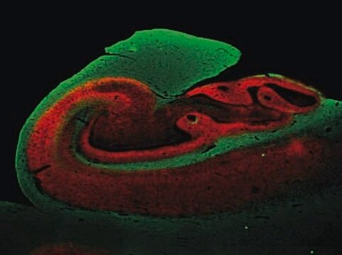
Immunohistochemistry Analysis: A representative lot detected a stronger synaptophysin immunoreactivity in the inner molecular layer of frozen dentate gyrus sections from a 3-week old monkey, while a stronger immunoreactivity was seen in the outer molecular layer of dentate gyrus sections from a 13-year old monkey (Lavenex, P., et al. (2007). Dev. Neurosci. 29(1-2):179–192).
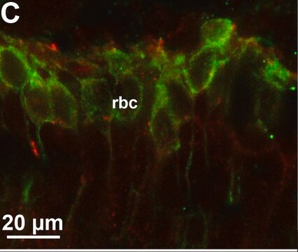
Immunohistochemistry Analysis: A representative lot immunostained nerve terminals at palisade ending using cat extraocular muscle (EOM) whole mount sections (Konakci, K.Z., et al. (2005). Invest. Ophthalmol. Vis. Sci. 46(1):155-165).
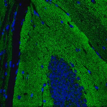
Immunohistochemistry Analysis: A representative lot detected synaptophysin immunoreactivity in human brain tissue sections (Honer, W.G., et al. (1993). Brain Res. 609(1-2):9-20).
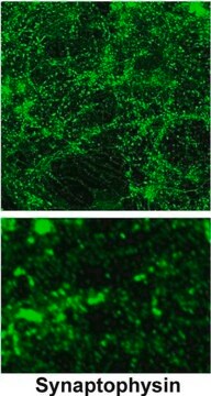
Immunocytochemistry Analysis: A representative lot detected punctate synatophysin immunoreactivity among cultured primary cerebrocortical neurons from rat embryos (Bragina, L., et al. (2006). J. Neurochem. 99(1):134-141.).
ELISA Analysis: Representative lots were employed as the capture antibody for the detection of synaptophysin in human temporal cortex, frontal cortex, and cerebellar cortex extracts by sandwich ELISA (Klucken, J., et al. (2006). Acta Neuropathol. 111(2):101-108; Fukumoto, H., et al. (2002). Arch. Neurol. 59(9):1381-1389).
Qualität
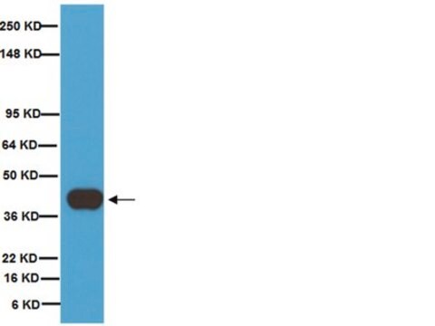
Western Blotting Analysis: 1.0 µg/mL of this antibody detected Synaptophysin in 10 µg of rat brain tissue lysate.
Zielbeschreibung
Physikalische Form
Lagerung und Haltbarkeit
Sonstige Hinweise
Haftungsausschluss
Sie haben nicht das passende Produkt gefunden?
Probieren Sie unser Produkt-Auswahlhilfe. aus.
Empfehlung
Lagerklassenschlüssel
12 - Non Combustible Liquids
WGK
WGK 2
Flammpunkt (°F)
Not applicable
Flammpunkt (°C)
Not applicable
Analysenzertifikate (COA)
Suchen Sie nach Analysenzertifikate (COA), indem Sie die Lot-/Chargennummer des Produkts eingeben. Lot- und Chargennummern sind auf dem Produktetikett hinter den Wörtern ‘Lot’ oder ‘Batch’ (Lot oder Charge) zu finden.
Besitzen Sie dieses Produkt bereits?
In der Dokumentenbibliothek finden Sie die Dokumentation zu den Produkten, die Sie kürzlich erworben haben.
Unser Team von Wissenschaftlern verfügt über Erfahrung in allen Forschungsbereichen einschließlich Life Science, Materialwissenschaften, chemischer Synthese, Chromatographie, Analytik und vielen mehr..
Setzen Sie sich mit dem technischen Dienst in Verbindung.