MAB1569
Anti-Microglia Antibody, clone 25F9
clone 25F9, Chemicon®, from mouse
Synonym(e):
Macrophages, Late State Inflammatory
About This Item
Empfohlene Produkte
Biologische Quelle
mouse
Qualitätsniveau
Antikörperform
purified immunoglobulin
Antikörper-Produkttyp
primary antibodies
Klon
25F9, monoclonal
Speziesreaktivität
human
Hersteller/Markenname
Chemicon®
Methode(n)
flow cytometry: suitable
immunocytochemistry: suitable
immunohistochemistry: suitable
Isotyp
IgG1
Versandbedingung
wet ice
Posttranslationale Modifikation Target
unmodified
Allgemeine Beschreibung
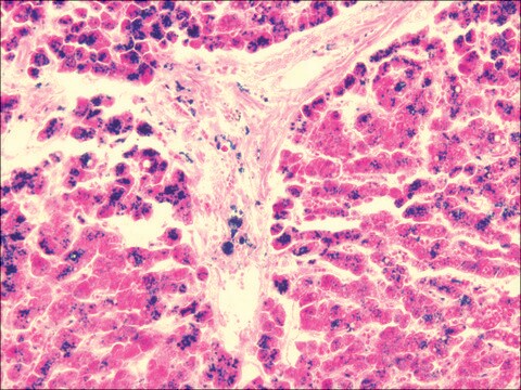
Isolated cells: Absent on freshly isolated monocytes and other blood cells; present on 40-50% of human monocytes after 6-7 day culture, also positive on some melanoma and carcinoma cell lines. Tissue sections: Kupffer cells, histiocytes (skin), macrophages of the thymus, in the germinal centers of lymph nodes and spleen, in mamma carcinoma, melanoma, osteocarcinoma and gastric cancer; excema, sarcoidosis, BCG granuloma; synovial lining cells, tuberculoid leprosy; no expression in lepramatous leprosy.
Spezifität
Antigen distribution: absent from freshly isolated monocytes and other blood cells; present on 40-50% of human monocytes after 6-7 days in culture, also positive on some melanoma and carcinoma lines. In tissue sections, clone identifies Kupffer cells, histiocytes (skin), macrophages of the thymus, in te germinal centers of lymph nodes and spleen, in mamma carcinoma, melanoma, osteocarcinoma and gastric cancer; excema, sarcoidosis, BCG granuloma;synovial lining cells, tuberculoid leprosy, however no expression in lepramatous leprosy.
Species reactivity is seen in human, rhesus monkey, and pig, other species not tested.
Anwendung
Immunocytochemistry
Optimal working dilutions must be determined by the end user.
Physikalische Form
Lagerung und Haltbarkeit
Rechtliche Hinweise
Sie haben nicht das passende Produkt gefunden?
Probieren Sie unser Produkt-Auswahlhilfe. aus.
Signalwort
Warning
H-Sätze
Gefahreneinstufungen
Acute Tox. 4 Dermal - Acute Tox. 4 Inhalation - Aquatic Chronic 3
Lagerklassenschlüssel
11 - Combustible Solids
WGK
WGK 3
Analysenzertifikate (COA)
Suchen Sie nach Analysenzertifikate (COA), indem Sie die Lot-/Chargennummer des Produkts eingeben. Lot- und Chargennummern sind auf dem Produktetikett hinter den Wörtern ‘Lot’ oder ‘Batch’ (Lot oder Charge) zu finden.
Besitzen Sie dieses Produkt bereits?
In der Dokumentenbibliothek finden Sie die Dokumentation zu den Produkten, die Sie kürzlich erworben haben.
Unser Team von Wissenschaftlern verfügt über Erfahrung in allen Forschungsbereichen einschließlich Life Science, Materialwissenschaften, chemischer Synthese, Chromatographie, Analytik und vielen mehr..
Setzen Sie sich mit dem technischen Dienst in Verbindung.


