
ROAHAHA
Roche
Anti-HA High Affinity
from rat IgG1
Synonym(s):
antibody
Sign Into View Organizational & Contract Pricing
All Photos(2)
About This Item
UNSPSC Code:
12352203
Recommended Products


biological source
rat
Quality Level
conjugate
unconjugated
antibody form
purified immunoglobulin
antibody product type
primary antibodies
clone
3F10, monoclonal
form
lyophilized
packaging
pkg of 50 μg (11867423001)
pkg of 500 μg (11867431001)
manufacturer/tradename
Roche
isotype
IgG1
epitope sequence
YPYDVPDYA
storage temp.
2-8°C
Related Categories
General description
Anti-HA High Affinity is a monoclonal antibody to the HA-peptide (clone 3F10). Anti-HA High Affinity recognizes the HA peptide sequence (YPYDVPDYA), derived from the influenza hemagglutinin protein. The antibody recognizes its antigenic determinant even when the HA peptide epitope is introduced into unrelated recombinant proteins by a technique known as "epitope tagging".
Specificity
Anti-HA, High Affinity recognizes the 9-amino acid sequence YPYDVPDYA, derived from the human influenza hemagglutinin (HA) protein. This epitope is also recognized in fusion proteins regardless of its position (N-terminal, C-terminal or internal).
Immunogen
Amino acids 98-106 from the human influenza virus hemagglutinin protein
Application
Use Anti-HA High Affinity for the detection of native influenza hemagglutinin protein and recombinant proteins that contain the HA epitope using:
- Dot blots
- ELISA
- Immunocytochemistry
- Immunoprecipitation
- Western blots
Quality
Function tested in western blot.
Preparation Note
Working concentration: Working concentration of conjugate depends on application and substrate.
The following concentrations should be taken as a guideline:
The following concentrations should be taken as a guideline:
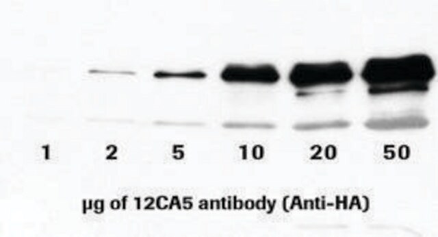
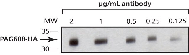
- ELISA: for detection 100 ng/ml; for coating 1 to 5 μg/ml
- Immunoprecipitation: 0.5 to 5 μg/ml
- Western blot: 50 to 200 ng/ml
Reconstitution
Add 0.5 ml (11 867 423 001; 50 μg) or 2.5 ml (11 867 431 001; 500 μg) double-distilled water to a final concentration of 100 μg/ml or 200 μg/ml respectively.
Rehydrate for 10 min prior to use.
Rehydrate for 10 min prior to use.
Other Notes
For life science research only. Not for use in diagnostic procedures.
Not finding the right product?
Try our Product Selector Tool.

Signal Word
Warning-Warning
Hazard Statements
Hazard Classifications
Aquatic Chronic 3 - Skin Sens. 1
Storage Class Code
11 - Combustible Solids
WGK
WGK 2
Flash Point(F)
does not flash
Flash Point(C)
does not flash
Choose from one of the most recent versions:
Already Own This Product?
Find documentation for the products that you have recently purchased in the Document Library.
Customers Also Viewed

Toshiki Kinuhata et al.
Journal of developmental biology, 10(4) (2022-11-23)
The first event of differentiation and morphogenesis in the optic vesicle (OV) is specification of the neural retina (NR) and retinal pigment epithelium (RPE), separating the inner and outer layers of the optic cup, respectively. Here, we focus on a
Elisabeth Stes et al.
Journal of proteome research, 13(6), 3107-3113 (2014-05-13)
Here, we apply the COmbined FRActional DIagonal Chromatography (COFRADIC) technology to enrich for ubiquitinated peptides and to identify sites of ubiquitination by mass spectrometry. Our technology bypasses the need to overexpress tagged variants of ubiquitin and the use of sequence-biased
Kristina Halbleib et al.
Molecular cell, 67(4), 673-684 (2017-07-12)
The unfolded protein response (UPR) is a conserved homeostatic program that is activated by misfolded proteins in the lumen of the endoplasmic reticulum (ER). Recently, it became evident that aberrant lipid compositions of the ER membrane, referred to as lipid bilayer stress
Alexandra Atienza-Manuel et al.
Development (Cambridge, England) (2021-11-06)
The vertebrate endocytic receptor CUBAM, consisting of three cubilin monomers complexed with a single amnionless molecule, plays a major role in protein reabsorption in the renal proximal tubule. Here, we show that Drosophila CUBAM is a tripartite complex composed of
Gizem Sancer et al.
Current biology : CB, 29(17), 2812-2825 (2019-08-14)
In the fly optic lobe, ∼800 highly stereotypical columnar microcircuits are arranged retinotopically to process visual information. Differences in cellular composition and synaptic connectivity within functionally specialized columns remain largely unknown. Here, we describe the cellular and synaptic architecture in
Our team of scientists has experience in all areas of research including Life Science, Material Science, Chemical Synthesis, Chromatography, Analytical and many others.
Contact Technical Service