MABS2046
Anti-ApoB Antibody, clone 2G11
clone 2G11, from mouse
Synonym(s):
Apolipoprotein B-100
Sign Into View Organizational & Contract Pricing
All Photos(1)
About This Item
UNSPSC Code:
12352203
eCl@ss:
32160702
NACRES:
NA.41
Recommended Products
biological source
mouse
Quality Level
antibody form
purified immunoglobulin
antibody product type
primary antibodies
clone
2G11, monoclonal
species reactivity
hamster, mouse, rat
should not react with
human
packaging
antibody small pack of 25 μg
technique(s)
ELISA: suitable
western blot: suitable
isotype
IgG1κ
NCBI accession no.
UniProt accession no.
shipped in
ambient
target post-translational modification
unmodified
Gene Information
mouse ... Apob(238055)
General description
Apolipoprotein B-100 (UniProt: E9Q414; also known as Apo B-100) is encoded by the Apob gene (Gene ID: 238055) in murine species. Apolipoprotein B is a major protein constituent of chylomicrons. Apo B particles are made up of lipids and proteins and each Apo B is bordered by a phospholipid monolayer with an inner core composed of variable amounts of triglycerides and cholesterol esters. Apo B is synthesized with a signal peptide (aa 1-27) that is subsequently cleaved off. Two isoforms of Apo B are produced- Apo B-100 is synthesized in the liver and Apo B-48 is produced in the intestine. Apo B-100 contains the region that binds to the LDL receptor. It contains VLDL particles, VLDL remnant particles and LDL particles. On the other hand, the Apo B-48 particles are made of chylomicrons and chylomicron remnant particles. Generally, LDL particles account for approximately 90% of Apo B particles in plasma. Abnormal trapping of an Apo B is a major factor in the development of atherosclerotic lesions. When Apo B particles degrade cholesterol is retained with macrophages that induce inflammatory response. Further entry and entrapment of Apo B can promote cardiovascular pathology over time. (Ref.: Sniderman, A et al. (2010). Nat. Rev. Endocrinol. 6(6); 335-346).
Specificity
Clone 2G11 detects Apolipoprotein B in Mouse, Rat, and Hamster plasma and tissues. It binds to ApoB-48 and ApoB-100, but not to ApoB-39.
Immunogen
ApoB-containing lipoproteins isolated from Apoe -/- Apob 48/48 mice.
Application
Detects Apolipoprotein B using this mouse monoclonal Anti-ApoB, clone 2G11 , Cat. No. MABS2046, is a mouse monoclonal antibody that detects Apolipoprotein B in Mouse, Rat, and Hamster and is tested for use in ELISA and Western Blotting.
Research Category
Signaling
Signaling
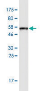
Western Blotting Analysis: 4 µg/mL from a representative lot detected ApoB in 10 µg of rat liver and mouse liver tissue lysate.
Western Blotting Analysis: A representative lot detected ApoB in Western Blotting applications (Nguyen, A.T., et. al. (2006). Biochem Biophys Acta. 1761(2):182-5; Cheng, D., et. al. (2016). J Biol Chem. 291(45):23793-23803).
ELISA Analysis: A representative lot detected ApoB in ELISA applications (Nguyen, A.T., et. al. (2006). Biochem Biophys Acta. 1761(2):182-5).
Quality
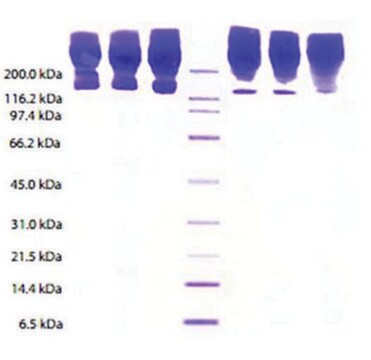
Evaluated by Western Blotting in rat serum.
Western Blotting Analysis: 4 µg/mL of this antibody detected ApoB in 10 µg of rat serum.
Western Blotting Analysis: 4 µg/mL of this antibody detected ApoB in 10 µg of rat serum.
Target description
~260 kDa observed; 509.43 kDa calculated. Uncharacterized bands may be observed in some lysate(s).
Physical form
Format: Purified
Protein G purified
Purified mouse monoclonal antibody IgG1 in buffer containing 0.1 M Tris-Glycine (pH 7.4), 150 mM NaCl with 0.05% sodium azide.
Storage and Stability
Stable for 1 year at 2-8°C from date of receipt.
Other Notes
Concentration: Please refer to lot specific datasheet.
Disclaimer
Unless otherwise stated in our catalog or other company documentation accompanying the product(s), our products are intended for research use only and are not to be used for any other purpose, which includes but is not limited to, unauthorized commercial uses, in vitro diagnostic uses, ex vivo or in vivo therapeutic uses or any type of consumption or application to humans or animals.
Not finding the right product?
Try our Product Selector Tool.
Certificates of Analysis (COA)
Search for Certificates of Analysis (COA) by entering the products Lot/Batch Number. Lot and Batch Numbers can be found on a product’s label following the words ‘Lot’ or ‘Batch’.
Already Own This Product?
Find documentation for the products that you have recently purchased in the Document Library.
Zsófia Onódi et al.
Frontiers in physiology, 9, 1479-1479 (2018-11-09)
Background: Extracellular vesicles (EVs) (isolated from blood plasma) are currently being extensively researched, both as biomarkers and for their therapeutic possibilities. One challenging aspect to this research is the efficient isolation of high-purity EVs from blood plasma in quantities sufficient
Masanori Fukushima et al.
The Journal of biological chemistry, 293(39), 15277-15289 (2018-08-25)
Extracellular vesicles are important carriers of cellular materials and have critical roles in cell-to-cell communication in both health and disease. Ceramides are implicated in extracellular vesicle biogenesis, yet the cellular machinery that mediates the formation of ceramide-enriched extracellular vesicles remains
Our team of scientists has experience in all areas of research including Life Science, Material Science, Chemical Synthesis, Chromatography, Analytical and many others.
Contact Technical Service


