Skip to Content
Merck
All Photos(2)

Key Documents

MABF244

Sigma-Aldrich

Anti-PAR-1, clone ATAP2 Antibody

clone ATAP2, from mouse

Synonym(s):

Proteinase-activated receptor 1, PAR-1, Coagulation factor II receptor, Thrombin receptor

Sign Into View Organizational & Contract Pricing


About This Item

UNSPSC Code:
12352203
eCl@ss:
32160702
NACRES:
NA.41
clone:
ATAP2, monoclonal
application:
FACS
ICC
IF
WB
species reactivity:
human, mouse
technique(s):
flow cytometry: suitable
immunocytochemistry: suitable
immunofluorescence: suitable
western blot: suitable
citations:
3

biological source

mouse

Quality Level

antibody form

purified immunoglobulin

antibody product type

primary antibodies

clone

ATAP2, monoclonal

species reactivity

human, mouse

technique(s)

flow cytometry: suitable
immunocytochemistry: suitable
immunofluorescence: suitable
western blot: suitable

isotype

IgG1κ

NCBI accession no.

UniProt accession no.

shipped in

wet ice

target post-translational modification

unmodified

Gene Information

human ... F2R(2149)

General description

The protein named Proteinase-activated receptor 1 (PAR-1/PAR1) or Coagulation factor II receptor, or Thrombin receptor and encoded by the human gene F2R/CR2R/PAR1/TR is a high affinity receptor for activated thrombin and part of the innate immune system response driven by proteinase-activated receptors (PARs). PARs are G protein-coupled receptors that transmit cellular responses to extracellular proteases and have important functions in vascular physiology, development, inflammation, and cancer progression. PAR1 is important for platelet activation and vascular development. PAR1 expression is limited to platelets and vascular endothelial cells and is localized to the cell membrane. Interestingly too, even the signal peptide of PAR1 (which guides the protein to the membrane) plays a role in cell signaling as if forms the intracellular angiogenesis inhibitor peptide Parstatin.

Immunogen

Linear peptide corresponding to human Par-1.

Application

Flow Cytometry Analysis: 4 µg from a representative lot detected Par-1 in 1X10E6 human thrombocytes (platelets).

Flow Cytometry Analysis: A representative lot from an independent laboratory detected Par-1 in CHRF-288 and HEL cells (Hoxie, J. A., et al. (1993). J Biol Chem. 268(18):13756-13763.).

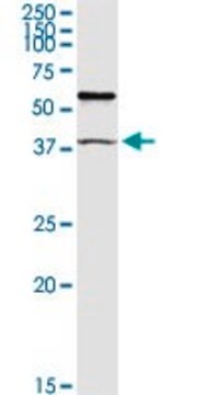
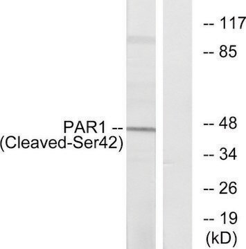
Western Blotting Analysis: A representative lot from an independent laboratory detected Par-1 in human platelet membrane cell lysate (Brass, L. F., et al. (1992). J Biol Chem. 267(20):13795-13798.).

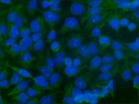
Immunocytochemistry Analysis: A representative lot from an independent laboratory detected Par-1 in CHRF-288 and HEL cells (Hoxie, J. A., et al. (1993). J Biol Chem. 268(18):13756-13763.).

Immunofluorescence Analysis: A representative lot from an independent laboratory detected Par-1 in CHRF-288 cells (Brass, L. F., et al. (1994). J Biol Chem. 269(4):2943-2952.).

This Anti-PAR-1 antibody, clone ATAP2 is validated for use in western blotting, flow cytometry, ICC & immunofluorescence for the detection of PAR-1.

Quality

Evaluated by Western Blotting in mouse lymph node tissue.

Western Blotting Analysis: 1 µg/mL of this antibody detected Par-1 in 10 µg of mouse lymph node tissue.

Target description

~50 kDa observed. Uncharacterized band(s) may be observed in some cell lysates.

Physical form

Format: Purified

Other Notes

Concentration: Please refer to the Certificate of Analysis for the lot-specific concentration.

Not finding the right product?  

Try our Product Selector Tool.

Storage Class Code

12 - Non Combustible Liquids

WGK

WGK 1

Flash Point(F)

Not applicable

Flash Point(C)

Not applicable


Certificates of Analysis (COA)

Search for Certificates of Analysis (COA) by entering the products Lot/Batch Number. Lot and Batch Numbers can be found on a product’s label following the words ‘Lot’ or ‘Batch’.

Already Own This Product?

Find documentation for the products that you have recently purchased in the Document Library.

Visit the Document Library

Internalization and recycling of activated thrombin receptors.
Hoxie, J A, et al.
The Journal of Biological Chemistry, 268, 13756-13763 (1993)
L F Brass et al.
The Journal of biological chemistry, 269(4), 2943-2952 (1994-01-28)
According to current models, human thrombin receptors are activated when thrombin cleaves the receptor's N terminus, exposing the tethered ligand domain, SFLLRN. In the megakaryoblastic CHRF-288 cell line, thrombin receptor activation is followed by the rapid internalization of > 90%
L F Brass et al.
The Journal of biological chemistry, 267(20), 13795-13798 (1992-07-25)
Based upon its recently cloned nucleotide sequence, the human platelet thrombin receptor is thought to be formed by a single polypeptide chain with seven transmembrane domains and an extracellular N terminus that can be cleaved by thrombin. As yet, however
Xiaofeng Cai et al.
Journal of thrombosis and haemostasis : JTH, 17(5), 803-817 (2019-03-14)
Essentials APC elicits cytoprotective responses in endothelial cells via EPCR-dependent cleavage of PAR1. APC inhibits LPS-mediated translocation and extracellular secretion of HMGB1 in endothelial cells. Signaling activity of APC inhibits LPS-mediated acetylation of HMGB1 by epigenetic mechanisms. APC inhibits LPS-mediated

Our team of scientists has experience in all areas of research including Life Science, Material Science, Chemical Synthesis, Chromatography, Analytical and many others.

Contact Technical Service