Saltar al contenido
MilliporeSigma

P1093

Sigma-Aldrich

Monoclonal Anti-Paxillin antibody produced in mouse

clone PXC-10, ascites fluid

Iniciar sesiónpara Ver la Fijación de precios por contrato y de la organización


About This Item

MDL number:
UNSPSC Code:
12352203
NACRES:
NA.41

biological source

mouse

conjugate

unconjugated

antibody form

ascites fluid

antibody product type

primary antibodies

clone

PXC-10, monoclonal

mol wt

antigen 68-40 kDa

contains

15 mM sodium azide

species reactivity

bovine, rat, human, chicken, hamster

technique(s)

immunocytochemistry: suitable
microarray: suitable
western blot: 1:500 using a cultured human cell line extract

isotype

IgG1

UniProt accession no.

shipped in

dry ice

storage temp.

−20°C

target post-translational modification

unmodified

Gene Information

human ... PXN(5829)
rat ... Pxn(360820)

General description

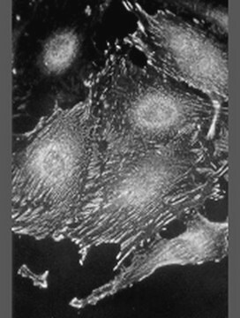
Monoclonal Anti-Paxillin (mouse IgG1 isotype) is derived from the PXC-10 hybridoma produced by the fusion of mouse myeloma cells and splenocytes from an immunized mouse. Paxillin (68 kDa), is a cytoskeletal component. It is found in the ends of actin stress fibers of the focal adhesions, but not in adherens junctions of the cells. The paxillin molecule has a single binding site for vinculin, and at least two binding sites for focal adhesion kinase (FAK), that are separated by an intervening sequence of 100 amino acids.

Specificity

Recognizes an epitope located in the LIM1 domain of the paxillin molecule.

Immunogen

C-terminal part of recombinant chicken paxillin (amino acids 305-559)

Application

Monoclonal Anti-Paxillin antibody produced in mouse has been used in immunoblotting and immunocytochemistry.

Biochem/physiol Actions

Paxillin interacts with several proteins including members of the src family of tyrosine kinases, the transforming protein v-crk, the cytoskeletal protein vinculin, and the tyrosine kinase, focal adhesion kinase (FAK). This interaction has suggested a function for paxillin as a molecular adaptor, responsible for the recruitment of structural and signaling molecules to focal adhesions.

Disclaimer

Unless otherwise stated in our catalog or other company documentation accompanying the product(s), our products are intended for research use only and are not to be used for any other purpose, which includes but is not limited to, unauthorized commercial uses, in vitro diagnostic uses, ex vivo or in vivo therapeutic uses or any type of consumption or application to humans or animals.

¿No encuentra el producto adecuado?  

Pruebe nuestro Herramienta de selección de productos.

Storage Class

10 - Combustible liquids

wgk_germany

WGK 3

flash_point_f

Not applicable

flash_point_c

Not applicable


Certificados de análisis (COA)

Busque Certificados de análisis (COA) introduciendo el número de lote del producto. Los números de lote se encuentran en la etiqueta del producto después de las palabras «Lot» o «Batch»

¿Ya tiene este producto?

Encuentre la documentación para los productos que ha comprado recientemente en la Biblioteca de documentos.

Visite la Librería de documentos

Paxillin: a new vinculin-binding protein present in focal adhesions.
Turner CE, et al.
The Journal of cell biology, 111(3), 1059-1068 (1990)
Paxillin: a crossroad in pathological cell migration
Lopez CAM, et al.
Journal of Hematology & Oncology, 10(1), 50-50 (2017)
Jung Yul Lim et al.
Biomaterials, 28(10), 1787-1797 (2007-01-16)
An important consideration in developing physical biomimetic cell-stimulating cues is that the in vivo extracellular milieu includes nanoscale topographic interfaces. We investigated nanoscale topography regulation of cell functions using human fetal osteoblastic (hFOB) cell culture on poly(l-lactic acid) and polystyrene
Gokhan Bahcecioglu et al.
International journal of biological macromolecules, 122, 1152-1162 (2018-09-16)
In this study, porcine fibrochondrocyte-seeded agarose, methacrylated gelatin (GelMA), methacrylated hyaluronic acid (MeHA) and GelMA-MeHA blend hydrogels, and 3D printed PCL scaffolds were tested under dynamic compression for potential meniscal regeneration in vitro. Cell-carrying hydrogels produced higher levels of extracellular
J L Funderburgh et al.
The Journal of biological chemistry, 276(47), 44173-44178 (2001-09-14)
Keratocytes of the corneal stroma secrete a unique population of proteoglycan molecules considered essential for corneal transparency. In healing corneal wounds, keratocytes exhibit a myofibroblastic phenotype in response to transforming growth factor beta (TGF-beta), characterized by expression of alpha-smooth muscle

Nuestro equipo de científicos tiene experiencia en todas las áreas de investigación: Ciencias de la vida, Ciencia de los materiales, Síntesis química, Cromatografía, Analítica y muchas otras.

Póngase en contacto con el Servicio técnico