Saltar al contenido
MilliporeSigma

M4276

Sigma-Aldrich

Monoclonal Anti-Myosin (Skeletal, Fast) antibody produced in mouse

clone MY-32, ascites fluid

Sinónimos:

Anti-Myosin Antibody

Iniciar sesiónpara Ver la Fijación de precios por contrato y de la organización


About This Item

MDL number:
UNSPSC Code:
12352203
NACRES:
NA.41

biological source

mouse

Quality Level

conjugate

unconjugated

antibody form

ascites fluid

antibody product type

primary antibodies

clone

MY-32, monoclonal

contains

15 mM sodium azide

species reactivity

rat, chicken, rabbit, mouse, human, bovine, guinea pig, feline

technique(s)

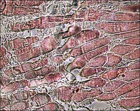
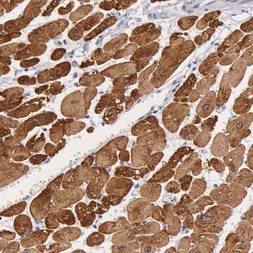
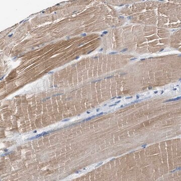
immunohistochemistry (formalin-fixed, paraffin-embedded sections): 1:400 using skeletal muscle tissue
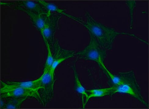
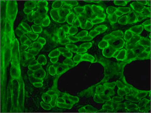
indirect immunofluorescence: 1:400
western blot: 1:1,000 using rabbit leg muscle extract

isotype

IgG1

UniProt accession no.

shipped in

dry ice

storage temp.

−20°C

target post-translational modification

unmodified

Gene Information

¿Está buscando productos similares? Visita Guía de comparación de productos

General description

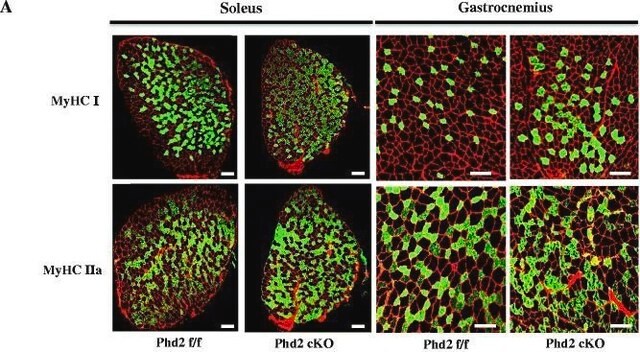
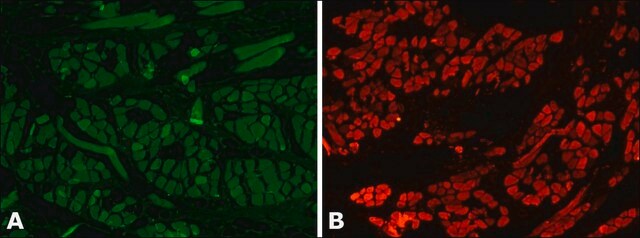
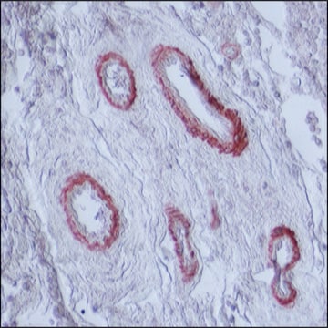
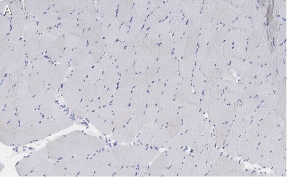
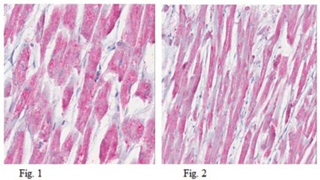
Localizes an epitope on the myosin heavy chain. Stains the fast (type II) and neonatal isomyosin molecules found in skeletal muscle, but does not stain cardiac muscle, smooth muscle or non-muscle myosin in cultured cells. Does react with human rhabdomyosarcomas.
Monoclonal Anti-Skeletal Myosin (mouse IgG1 isotype) is derived from the hybridoma produced by the fusion of mouse myeloma cells and splenocytes from an immunized mouse. Myosin is a 480,000 dalton protein known to interact with actin in muscle and in non-muscle cells. It contains two identical heavy chains (200,000 daltons each) and four light chains (15,000-26,000 daltons). Myosin molecules consist of two major regions: tails (rods) and heads; they aggregate into filaments through the tail region and interact with actin and with ATP through the head region. Multiple forms of myosin heavy chains exist for each muscle type-skeletal, cardiac, smooth and non-muscle isomyosin forms exist in different types of skeletal muscle, depending on the physiological function of the muscle. These are designated at type I (slow twitch) and type II (fast-twitch). Type II fibers can be further subdivided in types IIA, IIB, and IIC.

Specificity

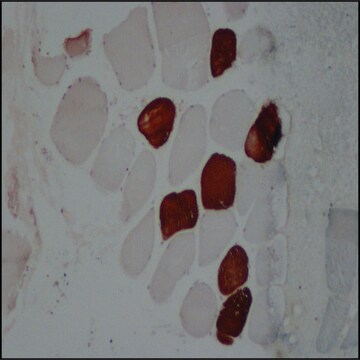
Monoclonal Anti-Skeletal Myosin is specific for the myosin heavy chain. It does not stain human or animal cardiac or smooth muscle myosin or cells grown by tissue culture (nonmuscle myosin). It has been demonstrated on human skeletal muscle that the antibody stains the fast twitch (type II) isomyosin molecules. Monoclonal Anti-Skeletal Myosin antibody does react with human rhabdomyosarcomas.

Immunogen

rabbit muscle myosin.

Application

Monoclonal Anti-Myosin (Skeletal, Fast) antibody produced in mouse has been used in:
  • immunohistochemistry
  • immunostaining
  • western blotting at a dilution 1:1000 and 1:90000†
  • indirect immunofluorescence (dilution 1:400) of formalin-fixed, paraffin-embedded sections of human or animal skeletal muscle tissue preparation.
  • dot immunobinding on muscle extracts or purified myosin preparations
Monoclonal Anti-Skeletal Myosin may be used for staining of human, rabbit, rat, mouse, bovine, chicken and guinea pig skeletal myosin. Monoclonal Anti-Skeletal Myosin antibody to fast-twitch skeletal myosin may be used for detecting cross striated muscle differentiation in tumors. The antibody localizes an epitope on the myosin chain that is stable to the routine formalin-fixation and paraffin-embedding process.
The level of mysosin (fast) in serum samples from sportsmen with past injury was determined by western blot using monoclonal mouse anti-myosin (skeletal/fast) as the primary antibody at a dilution of 1:90000.

Disclaimer

Unless otherwise stated in our catalog or other company documentation accompanying the product(s), our products are intended for research use only and are not to be used for any other purpose, which includes but is not limited to, unauthorized commercial uses, in vitro diagnostic uses, ex vivo or in vivo therapeutic uses or any type of consumption or application to humans or animals.

¿No encuentra el producto adecuado?  

Pruebe nuestro Herramienta de selección de productos.

Storage Class

10 - Combustible liquids

wgk_germany

WGK 3

flash_point_f

Not applicable

flash_point_c

Not applicable


Certificados de análisis (COA)

Busque Certificados de análisis (COA) introduciendo el número de lote del producto. Los números de lote se encuentran en la etiqueta del producto después de las palabras «Lot» o «Batch»

¿Ya tiene este producto?

Encuentre la documentación para los productos que ha comprado recientemente en la Biblioteca de documentos.

Visite la Librería de documentos

An optimized histochemical method to assess skeletal muscle glycogen and lipid stores reveals two metabolically distinct populations of type I muscle fibers
Prats C, et al.
PLoS ONE, 8(10), e77774-e77774 (2013)
Quantitative Human Physiology: An Introduction (2012)
Bleeding efficiency, microbiological quality and oxidative stability of meat from goats subjected to slaughter without stunning in comparison with different methods of pre-slaughter electrical stunning
Sabow AB, et al.
Testing, 11(4), e0152661-e0152661 (2016)
Contractility and kinetics of human fetal and human adult skeletal muscle
Racca AW, et al.
The Journal of Physiology, 591(12), 3049-3061 (2013)
Espen E Spangenburg et al.
Journal of biomedicine & biotechnology, 2011, 598358-598358 (2011-10-01)
Triglyceride storage is altered across various chronic health conditions necessitating various techniques to visualize and quantify lipid droplets (LDs). Here, we describe the utilization of the BODIPY (493/503) dye in skeletal muscle as a means to analyze LDs. We found

Nuestro equipo de científicos tiene experiencia en todas las áreas de investigación: Ciencias de la vida, Ciencia de los materiales, Síntesis química, Cromatografía, Analítica y muchas otras.

Póngase en contacto con el Servicio técnico