Saltar al contenido
MilliporeSigma

L8271

Sigma-Aldrich

Monoclonal Anti-Laminin antibody produced in mouse

clone LAM-89, ascites fluid

Sinónimos:

Anti-Laminin Antibody

Iniciar sesiónpara Ver la Fijación de precios por contrato y de la organización


About This Item

MDL number:
UNSPSC Code:
12352203
NACRES:
NA.41

biological source

mouse

Quality Level

conjugate

unconjugated

antibody form

ascites fluid

antibody product type

primary antibodies

clone

LAM-89, monoclonal

contains

15 mM sodium azide

species reactivity

feline, human, pig

should not react with

rabbit, lizard, sheep, carp, canine, chicken, rat, guinea pig, goat, frog, snake

technique(s)

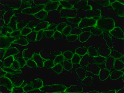
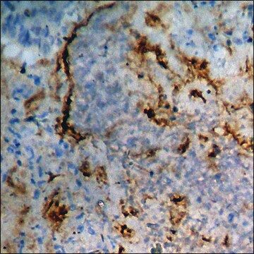
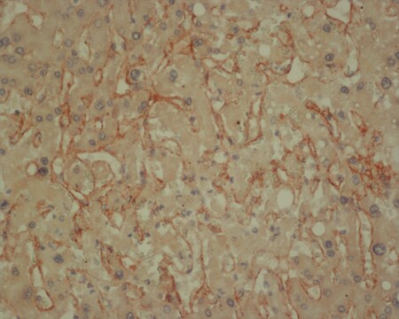
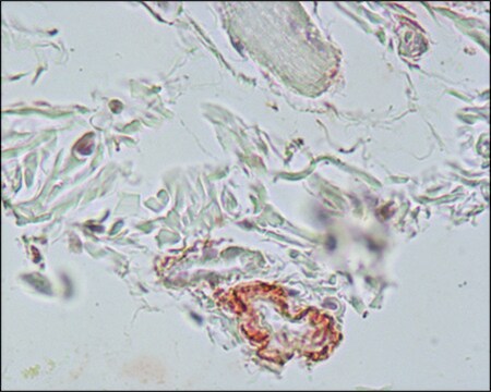
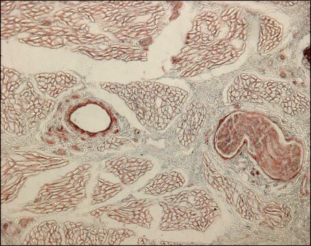
immunohistochemistry (formalin-fixed, paraffin-embedded sections): 1:1,000 using enzyme-treated human tissue sections
indirect ELISA: suitable
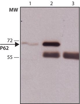
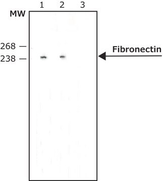
western blot: suitable

isotype

IgG1

UniProt accession no.

shipped in

dry ice

storage temp.

−20°C

target post-translational modification

unmodified

General description

Laminin, a 900kD (approx.) complex extracellular glycoprotein, is the most abundant structural and biologically active component present in the basement membranes. It is a cell substrate adhesion protein and thus has a crucial role in promoting neurite regeneration and modulating cellular functions like differentiation, cell shape and cell movement. Monoclonal Anti-laminin antibody can be used in immunohistochemistry to classify various disease processes involving basement membranes, to mark blood vessel walls of different species and to determine the origin and classification of human tumours. It can also be used in dot blot assays. Mouse anti- laminin antibody reacts specifically with purified human laminin but not with collagen IV, fibronectin, vitronectin or chondroitin sulfate types A, B, and C.
Monoclonal Anti-Laminin (mouse IgG1 isotype) is derived from the hybridoma produced by the fusion of mouse myeloma cells and splenocytes from an immunized mouse. Laminin, the most abundant structural and biologically active component in basement membranes, is a complex extracellular glycoprotein with an approximate molecular weight of 900 kDa. Laminin is composed of one A chain (400 kDa) one B1 chain (215 kDa) and one B2 chain (205 kDa) all held together by disulfide bonds. Laminin is only found in significant quantities in basement membranes, the thin extracellular matrices that surround epithelial tissue, nerve, fat cells and smooth, striated and cardiac muscle.

Immunogen

human laminin.

Application

Monoclonal Anti-Laminin antibody produced in mouse has been used in:
  • immunohistochemistry to stain matrix proteins
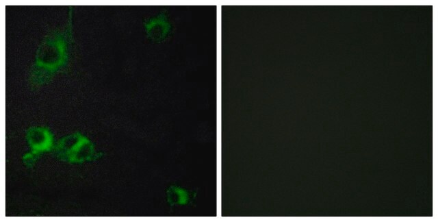
  • double immunofluorescence staining
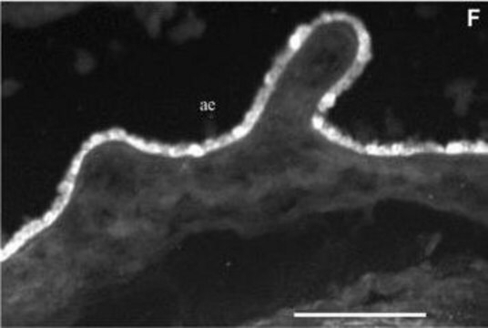
  • indirect immunofluorescence
  • immunogold labelling for electron microscopy

Monoclonal Anti-laminin antibody can be used in indirect immunostaining to determine the distribution of laminin. It may also be used for ELISA, Immunofluorescence and western blotting.

Biochem/physiol Actions

Laminin is a cell substrate adhesion protein and thus has a crucial role in promoting neurite regeneration and modulating cellular functions like differentiation, cell shape and cell movement because it appears to be an important cell substrate adhesion protein. It plays an important role in many aspects of the cell biology. Variations in the expression of this protein have been observed in embryogenesis, organogenesis, post traumatic healing and cancer.

Disclaimer

Unless otherwise stated in our catalog or other company documentation accompanying the product(s), our products are intended for research use only and are not to be used for any other purpose, which includes but is not limited to, unauthorized commercial uses, in vitro diagnostic uses, ex vivo or in vivo therapeutic uses or any type of consumption or application to humans or animals.

¿No encuentra el producto adecuado?  

Pruebe nuestro Herramienta de selección de productos.

Storage Class

12 - Non Combustible Liquids

wgk_germany

nwg

flash_point_f

Not applicable

flash_point_c

Not applicable


Certificados de análisis (COA)

Busque Certificados de análisis (COA) introduciendo el número de lote del producto. Los números de lote se encuentran en la etiqueta del producto después de las palabras «Lot» o «Batch»

¿Ya tiene este producto?

Encuentre la documentación para los productos que ha comprado recientemente en la Biblioteca de documentos.

Visite la Librería de documentos

Bryon R McKay et al.
The Journal of physiology, 588(Pt 17), 3307-3320 (2010-07-14)
In humans, muscle satellite cell (SC) enumeration is an important measurement used to determine the myogenic response to various stimuli. To date, the standard practice for enumeration is immunohistochemistry (IHC) using antibodies against common SC markers (Pax7, NCAM). Flow cytometry
Activation of hepatocyte growth factor receptor, c-met, in renal tubules is required for renoprotection after acute kidney injury
Zhou D, et al.
Kidney International, 84(3), 509-520 (2013)
Martin F Gerrits et al.
Journal of lipid research, 51(8), 2394-2404 (2010-03-25)
Inter-individual variability in weight gain and loss under energy surfeit and deficit conditions, respectively, are well recognized but poorly understood phenomena. We documented weight loss variability in an intensively supervised clinical weight loss program and assessed skeletal muscle gene expression
Laminin biosynthesis in the extracellular matrix-producing cell line PFHR9 studied with monoclonal and polyclonal antibodies
Lissitzky JC, et al.
The Biochemical Journal, 250(3), 843-852 (1988)
Jack A Helliwell et al.
Journal of tissue engineering, 8, 2041731417724011-2041731417724011 (2017-08-18)
The aim of this study was to adapt a proprietary decellularisation process for human dermis for use with porcine skin. Porcine skin was subject to: sodium chloride (1 M) to detach the epidermis, trypsin paste to remove hair follicles, peracetic acid

Nuestro equipo de científicos tiene experiencia en todas las áreas de investigación: Ciencias de la vida, Ciencia de los materiales, Síntesis química, Cromatografía, Analítica y muchas otras.

Póngase en contacto con el Servicio técnico