CLL1135
SKOV3 Cells GFP-HER2
human female ovary (Source Disease: Ovarian adenocarcinoma)
Iniciar sesiónpara Ver la Fijación de precios por contrato y de la organización
About This Item
UNSPSC Code:
41106514
NACRES:
NA.81
Productos recomendados
Nombre del producto
SKOV3 Cells GFP-HER2,
biological source
human female ovary (Source Disease: Ovarian adenocarcinoma)
Quality Level
OMIM accession no.
storage temp.
−196°C
Gene Information
human ... ERBB2(2064) , HER2(2064)
General description
SKOV3 GFP-HER2 are ovarian adenocarcinoma cells from a human 64 year old caucasion female having a ZFN modification creating a HER2-GFP transgene expressed from the endogenous HER2 gene locus.
This cell line was derived from ATCC Catalog No. HTB-77.
This cell line was derived from ATCC Catalog No. HTB-77.
Application
SKOV3 Cells GFP-HER2 has been used for live-cell screening assays to find compounds that affect cell structure or signaling pathways.
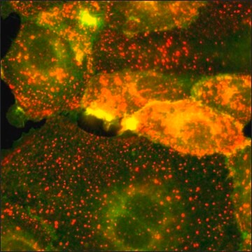
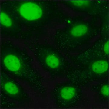
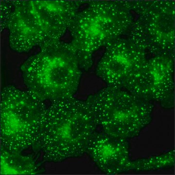
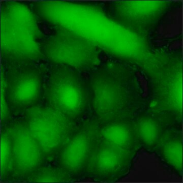
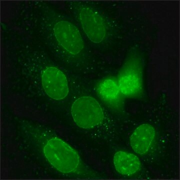
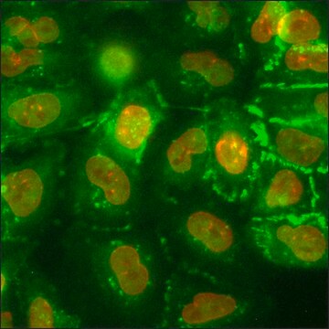
This product is a human SKOV3 cell line in which the genomic HER2 gene has been endogenously tagged with a Green Fluorescent Protein (GFP) gene using CompoZr® Zinc Finger Nuclease technology. Integration resulted in endogenous expression of the fusion protein in which GFP is attached to the C-terminus of HER2. Fluorescence imaging shows characteristic HER2 membrane expression. This stable cell line was expanded from a single clone. The target′s gene regulation and corresponding protein function are preserved in contrast to cell lines with overexpression via an exogenous promoter.
To learn more, please visit the Cellular Reporter Cell Line webpage
To learn more, please visit the Cellular Reporter Cell Line webpage
Biochem/physiol Actions
Human epidermal growth factor receptor 2 (HER2) signaling pathway promotes cell proliferation and survival in majority of breast cancers. Thus, overexpression of this protein leads to breast cancer. Heterodimeric complex of HER2 and phosphatidylinositide 3-kinase (PI3K) is the most potent stimulator of the phosphatidylinositol-3-kinase (PI3K)/Akt anti-apoptosis pathway. Upregulated expression of HER2 is associated with the development of ovarian, colorectal, pancreatic, endometrial and gastric cancers. Trastuzumab, an antibody, works by binding to a domain in the external domain of HER2. This domain is missing in p95, a truncated form of HER2, and hence these cancer cells show resistance to trastuzumab. HER2 protein can be used as a prognostic marker and as a therapeutic option for gynecologic cancers.
Features and Benefits
Zinc Finger Nuclease (ZFN)-mediated targeted integration of a fluorescent GFP tag into the last exon of the HER2 gene on chromosome 17q21.1 to create a cell line exhibiting stable expression of the transgene tagged with GFP on the C-terminus of the protein.
The SKOV3 cells are adherent, with a doubling time of approx. 48 hours.
The SKOV3 cells are adherent, with a doubling time of approx. 48 hours.
Quality
Tested for Mycoplasma, sterility, post-freeze viability, short terminal repeat (STR) analysis for cell line identification, cytochrome oxidase I (COI) analysis for cell line species confirmation.
Preparation Note
Media Renewal changes two to three times per week.
Rapidly thaw vial by gentle agitation in 37°C water bath (~2 minutes), keeping vial cap out of the water. Decontaminate with 70% ethanol, add 9 mL culture media and centrifuge 125 x g (5-7 minutes). Resuspend in complete culture media and incubate at 37°C in a 5% CO2 atmosphere.
Subculture Ratio: approx. 1:3-1:6
The base medium for this cell line is McCoy′s 5A Medium, Cat. No. M8403. To make the complete growth medium, add the following components to the base medium: fetal bovine serum, Cat. No. F2442, to a final concentration (v/v) of 10% and L-glutamine, Cat. No. G7513, at a final concentration of 1.5 mM.
Cell freezing medium-DMSO 1X, Cat. No. C6164.
Legal Information
CompoZr is a registered trademark of Merck KGaA, Darmstadt, Germany
Disclaimer
RESEARCH USE ONLY. This product is regulated in France when intended to be used for scientific purposes, including for import and export activities (Article L 1211-1 paragraph 2 of the Public Health Code). The purchaser (i.e. enduser) is required to obtain an import authorization from the France Ministry of Research referred in the Article L1245-5-1 II. of Public Health Code. By ordering this product, you are confirming that you have obtained the proper import authorization.
Storage Class
10 - Combustible liquids
wgk_germany
WGK 3
flash_point_f
188.6 °F - closed cup
flash_point_c
87 °C - closed cup
Elija entre una de las versiones más recientes:
Certificados de análisis (COA)
Lot/Batch Number
¿No ve la versión correcta?
Si necesita una versión concreta, puede buscar un certificado específico por el número de lote.
¿Ya tiene este producto?
Encuentre la documentación para los productos que ha comprado recientemente en la Biblioteca de documentos.
HER2 expression beyond breast cancer: therapeutic implications for gynecologic malignancies.
English DP, et al.
Molecular Diagnosis & Therapy, 17(2), 85-99 (2013)
The role of p95HER2 in trastuzumab resistance in breast cancer.
Ozkavruk Eliyatkin N, et al.
Journal of B.U.ON. : Official Journal of the Balkan Union of Oncology, 21(2), 382-389 (2016)
HER2: biology, detection, and clinical implications.
Gutierrez C and Schiff R.
Archives of Pathology & Laboratory Medicine, 135(1), 55-62 (2011)
Nuestro equipo de científicos tiene experiencia en todas las áreas de investigación: Ciencias de la vida, Ciencia de los materiales, Síntesis química, Cromatografía, Analítica y muchas otras.
Póngase en contacto con el Servicio técnico