Saltar al contenido
MilliporeSigma
Todas las fotos(1)

Documentos clave

236A-1

Sigma-Aldrich

CEA Rabbit Polyclonal Antibody

Iniciar sesiónpara Ver la Fijación de precios por contrato y de la organización


About This Item

UNSPSC Code:
12352203
NACRES:
NA.41

biological source

rabbit

Quality Level

100
500

conjugate

unconjugated

antibody form

Ig fraction of antiserum

antibody product type

primary antibodies

clone

polyclonal

description

For In Vitro Diagnostic Use in Select Regions (See Chart)

form

buffered aqueous solution

species reactivity

human

packaging

vial of 0.1 mL concentrate (236A-14)
vial of 0.5 mL concentrate (236A-15)
bottle of 1.0 mL predilute (236A-17)
vial of 1.0 mL concentrate (236A-16)
bottle of 7.0 mL predilute (236A-18)

manufacturer/tradename

Cell Marque

technique(s)

immunohistochemistry (formalin-fixed, paraffin-embedded sections): 1:200-1:500

control

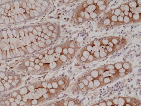
colon

shipped in

wet ice

storage temp.

2-8°C

visualization

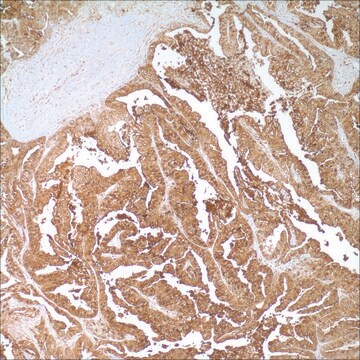
cytoplasmic

Gene Information

human ... CEACAM5(1048)

General description

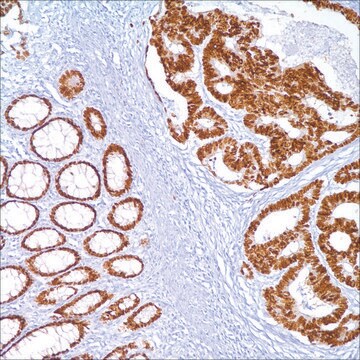
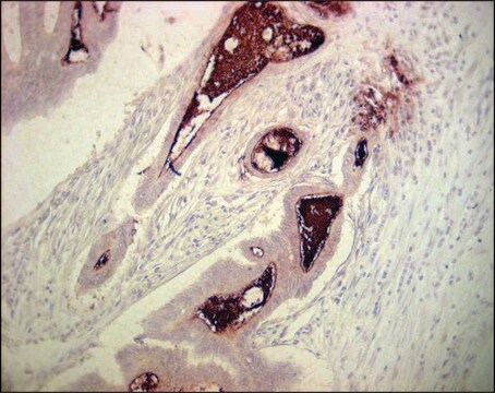
Anti-CEA is employed essentially as a tool to assist in the distinction between adenocarcinoma and epithelioid malignant mesotheliomas, along with other markers such as those against calretinin, CK 5&6, CD15, HBME-1, MOC-31, and Ber-EP4. Another suggested use of anti-CEA is to immunophenotype various metastatic adenocarcinomas as a means of identifying their origin within a panel of different markers. Anti-CEA positivity is seen in adenocarcinomas from the lung, colon, stomach, esophagus, pancreas, gallbadder, urachus, salivary gland, ovary, and endocervix. Polyclonal anti-CEA is useful in staining hepatocellular carcinoma in a canalicular pattern.

Linkage

CEA Positive Control Slides, Product No. 236S, are available for immunohistochemistry (formalin-fixed, paraffin-embedded sections).

Physical form

Solution in Tris Buffer, pH 7.3-7.7, with 1% BSA and <0.1% Sodium Azide

Preparation Note

Download the IFU specific to your product lot and formatNote: This requires a keycode which can be found on your packaging or product label.

Other Notes

For Technical Service please contact: 800-665-7284 or email: service@cellmarque.com

Legal Information

Cell Marque is a trademark of Merck KGaA, Darmstadt, Germany

¿No encuentra el producto adecuado?  

Pruebe nuestro Herramienta de selección de productos.


Elija entre una de las versiones más recientes:

Certificados de análisis (COA)

Lot/Batch Number

¿No ve la versión correcta?

Si necesita una versión concreta, puede buscar un certificado específico por el número de lote.

¿Ya tiene este producto?

Encuentre la documentación para los productos que ha comprado recientemente en la Biblioteca de documentos.

Visite la Librería de documentos

A Alkushi et al.
Virchows Archiv : an international journal of pathology, 442(3), 271-277 (2003-03-21)
Adenocarcinomas of the uterine cervix show a wide range of morphological features, and can be confused with endometrial adenocarcinoma in biopsy or curetting specimens. The objective of this study was to use tissue microarray technology to evaluate the immunoprofile of
Carl Morrison et al.
Modern pathology : an official journal of the United States and Canadian Academy of Pathology, Inc, 15(12), 1279-1287 (2002-12-14)
The distinction of hepatocellular carcinoma (HCC) from metastatic adenocarcinoma (MA) and cholangiocarcinoma (CC) in some cases requires the use of immunohistochemistry. CD10 has recently been suggested as a useful stain for HCC. We directly compared CD10 with other immunohistochemical markers
K Sheahan et al.
American journal of clinical pathology, 94(2), 157-164 (1990-08-01)
To evaluate the role of carcinoembryonic antigen (CEA) in solving problems of tumor histogenesis in surgical pathology, monoclonal antibodies to four distinct epitopes of CEA (E-Z-EM) were applied to paraffin sections of 303 epithelial neoplasms from multiple sites. Two epitopes
P W Shield et al.
American journal of clinical pathology, 105(2), 157-162 (1996-02-01)
The contribution of immunocytochemical (ICC) staining to the diagnosis of a range of cytologic specimens reported over a 20-month period was retrospectively assessed. A total of 194 cases (1.6% of total workload) were stained for diagnostic purposes in this period.

Nuestro equipo de científicos tiene experiencia en todas las áreas de investigación: Ciencias de la vida, Ciencia de los materiales, Síntesis química, Cromatografía, Analítica y muchas otras.

Póngase en contacto con el Servicio técnico