Saltar al contenido
MilliporeSigma
Todas las fotos(2)

Documentos clave

MABS82

Sigma-Aldrich

Anti-phospho-p70 S6 Kinase (Thr389) Antibody, clone 10G7.1

clone 10G7.1, from mouse

Sinónimos:

Ribosomal protein S6 kinase beta-1, S6K-beta-1, S6K1, 70 kDa ribosomal protein S6 kinase 1, Short name=P70S6K1, p70-S6K 1, Ribosomal protein S6 kinase I, Serine/threonine-protein kinase 14A, p70 ribosomal S6 kinase alpha, p70 S6 kinase alpha, p70 S6K-alp

Iniciar sesiónpara Ver la Fijación de precios por contrato y de la organización


About This Item

UNSPSC Code:
12352203
eCl@ss:
32160702
NACRES:
NA.41

biological source

mouse

Quality Level

antibody form

purified antibody

antibody product type

primary antibodies

clone

10G7.1, monoclonal

species reactivity

human, mouse

technique(s)

immunocytochemistry: suitable
western blot: suitable

isotype

IgG2aκ

NCBI accession no.

UniProt accession no.

shipped in

wet ice

target post-translational modification

phosphorylation (pThr389)

Gene Information

human ... RPS6KB1(6198)

General description

The p70 S6 kinase is a ubiquitous cytoplasmic protein that is activated in response to cytokines, and growth factors including the epidermal growth factor and fibroblast growth factor. It lies downstream of the mTOR/PI3K pathway and is phosphorylated on multiple residues: Thr389, Thr229, Thr421, Ser371, Ser404, Ser411, Ser418, and Ser424. Thr389, Ser371, and Ser404 are located in the linker domain; whereas Thr421, Ser411, Ser418, Ser424 are located in the autoinhibitory domain, and Thr229 is found in the catalytic domain. These sites may be phosphorylated in a hierarchical sequence leading to the activation of the p70 S6 kinase. The p70 S6 kinase phosphorylates the S6 protein on the 40S ribosomal protein (rpS6), and plays a role in multiple processes including regulation of mRNA molecules with 5’-terminal oligopyrimidine tracts, protein synthesis, cell growth, proliferation, and cell survival. The activity of this kinase may be important in diabetes, cancer, and aging.

Specificity

This antibody recognizes p70 S6 Kinase phosphorylated at (Thr389). This antibody also recognizes isoforms Alpha I and Alpha II.

Immunogen

Epitope: Phosphorylated (Thr389)
KLH-conjugated linear peptide corresponding to mouse p70 S6 Kinase phosphorylated at (Thr389).

Application

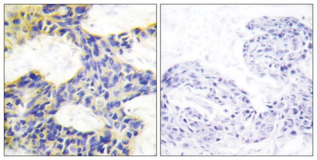
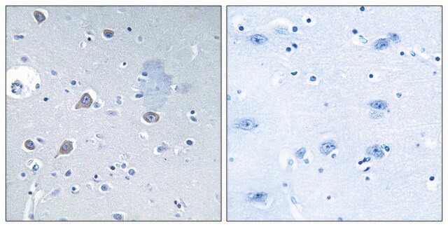
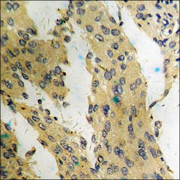
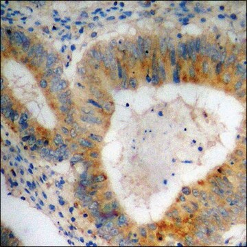
Immunocytochemistry Analysis: A 1:500 dilution from a representative lot detected p70 S6 Kinase in A431 cells.
Research Category
Signaling
Research Sub Category
Cell Cycle, DNA Replication & Repair
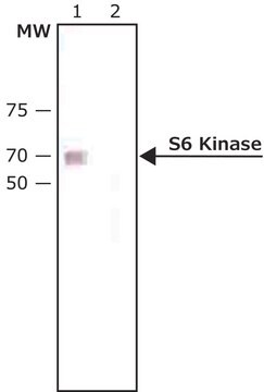
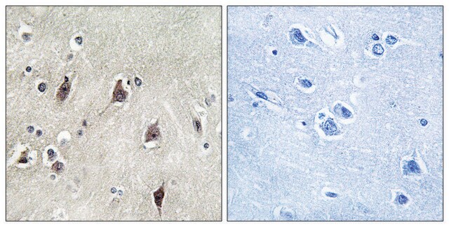
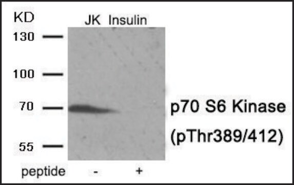
This Anti-phospho-p70 S6 Kinase (Thr389) Antibody, clone 10G7.1 is validated for use in Western Blotting, ICC for the detection of phospho-p70 S6 Kinase (Thr389).

Quality

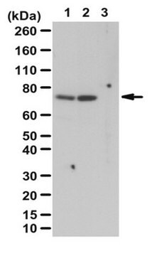
Evaluated by Western Blot in untreated and serum treated NIH/3T3 cell lysates.

Western Blot Analysis: 1 µg/mL of this antibody detected p70 S6 Kinase on 10 µg of untreated and serum treated NIH/3T3 cell lysates.

Target description

~70/85 kDa observed. This protein recognizes isoforms Alpha I and Alpha II. Uncharacterized bands appear at ~18 and 200 kDa in some lysates depending on treatment conditions.

Linkage

Replaces: 04-392

Physical form

Format: Purified
Protein G Purified
Purified mouse monoclonal IgG2aκ in buffer containing 0.1 M Tris-Glycine (pH 7.4), 150 mM NaCl with 0.05% sodium azide.

Storage and Stability

Stable for 1 year at 2-8°C from date of receipt.

Analysis Note

Control
Untreated and serum treated NIH/3T3 cell lysates

Other Notes

Concentration: Please refer to the Certificate of Analysis for the lot-specific concentration.

Disclaimer

Unless otherwise stated in our catalog or other company documentation accompanying the product(s), our products are intended for research use only and are not to be used for any other purpose, which includes but is not limited to, unauthorized commercial uses, in vitro diagnostic uses, ex vivo or in vivo therapeutic uses or any type of consumption or application to humans or animals.

¿No encuentra el producto adecuado?  

Pruebe nuestro Herramienta de selección de productos.

Storage Class

12 - Non Combustible Liquids

wgk_germany

WGK 1

flash_point_f

Not applicable

flash_point_c

Not applicable


Certificados de análisis (COA)

Busque Certificados de análisis (COA) introduciendo el número de lote del producto. Los números de lote se encuentran en la etiqueta del producto después de las palabras «Lot» o «Batch»

¿Ya tiene este producto?

Encuentre la documentación para los productos que ha comprado recientemente en la Biblioteca de documentos.

Visite la Librería de documentos

Yu-Ting Kang et al.
Oncotarget, 8(62), 105536-105552 (2017-12-30)
Autophagy is an intracellular recycling and degradation process for regulating tumor progression, survival and drug resistance. Nickel compounds have been identified as human carcinogens. However, the role of nickel-induced autophagy in lung carcinogenesis has not yet been fully elucidated. In
Jin-Zhu Wang et al.
The Journal of clinical investigation, 132(8) (2022-03-04)
Y-box-binding protein 1 (YB-1) is a multifunctional RNA binding protein involved in virtually every step of RNA metabolism. However, the functions and mechanisms of YB-1 in one of the most aggressive cancers, glioblastoma, are not well understood. In this study
Kristin Dauner et al.
The Journal of biological chemistry, 292(14), 5737-5747 (2017-02-16)
The plasma membrane of mammalian cells undergoes constitutive endocytosis, endocytic sorting, and recycling, which delivers nutrients to the lysosomes. The receptors, along with membrane lipids, are normally returned to the plasma membrane to sustain this action. It is not known
Tingting Sun et al.
Aging, 12(23), 24208-24218 (2020-11-24)
The 5-year survival rate of ovarian cancer patients is only 47%, and developing novel drugs for ovarian cancer is needed. Herein, we evaluated if and how SRT2183, a sirtuin-1 activator, impairs the ovarian cancer cells. OVCAR-3 and A2780 cells were

Nuestro equipo de científicos tiene experiencia en todas las áreas de investigación: Ciencias de la vida, Ciencia de los materiales, Síntesis química, Cromatografía, Analítica y muchas otras.

Póngase en contacto con el Servicio técnico