Saltar al contenido
MilliporeSigma
Todas las fotos(1)

Documentos

MABF957

Sigma-Aldrich

Anti-CLEC-2 Antibody, clone AYP1

clone AYP1, from mouse

Sinónimos:

C-type lectin domain family 1 member B, C-type lectin-like receptor 2, CLEC-2

Iniciar sesiónpara Ver la Fijación de precios por contrato y de la organización


About This Item

UNSPSC Code:
12352203
eCl@ss:
32160702
NACRES:
NA.43

biological source

mouse

Quality Level

antibody form

purified immunoglobulin

antibody product type

primary antibodies

clone

AYP1, monoclonal

species reactivity

human

technique(s)

flow cytometry: suitable

isotype

IgG1κ

NCBI accession no.

UniProt accession no.

shipped in

ambient

target post-translational modification

unmodified

Gene Information

human ... CLEC1B(51266)

General description

C-type lectin domain family 1 member B (UniProt Q9P126; also known as C-type lectin-like receptor 2, CLEC-2) is encoded by the CLEC1B (also known as CLEC2, UNQ721/PRO1384) gene (Gene ID 51266) in human. CLEC-2 is a type II single-transmembrane (a.a. 34-54) protein with a large extracellular (a.a. 55-229) region that contains a C-type lectin domain (a.a. 109-217) and a short N-terminal cytoplasmic tail (a.a. 1-33) with a single hem-immunoreceptor tyrosine-based activation motif (hemITAM; a.a. 7-10). CLEC-2 expression is restricted to platelets, where it mediates platelets activation via its hemITAM motif, leading to proteolytic cleavage of two other platelet ITAM receptors, glycoprotein (GP)VI and Fc RIIa, but not of CLEC-2 itself. CLEC-2 functions as the receptor for the type I transmembrane GP podoplanin that is widely expressed outside of the vasculature, including lymphatic endothelial cells, type 1 lung alveolar cells, lymph node stromal cells, the choroid plexus epithelium, inflammatory macrophages, as well as a subset of activated T-helper (Th)17 cells. Patients with rheumatoid arthritis show increased plasma levels of CLEC-2-positive microparticles. These microparticles are derived from activated platelets and are negative for GPVI, while microparticles in healthy donors are predominantly derived from megakaryocytes and are positive for both CLEC-2 and GPVI. Platelet-specific deletion of CLEC-2 or one of its downstream signaling proteins, Syk, SLP-76, or PLC 2, leads to a number of developmental problems in mice, including blood-lymphatic mixing in midgestation.

Specificity

Clone AYP1 targets an extracellular epitope present in both spliced isoforms of human CLEC-2/CLEC1B reported by UniProt (Q9P126).

Immunogen

His-tagged recombinant human CLEC-2 extracellular fragment.

Application

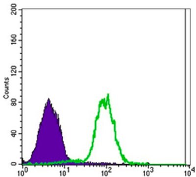
Flow Cytometry Analysis: A representative lot detected doxycycline-dependent expression induction of exogenously transfected CLEC-2/CLEC1B using T-REx 293 cells (Gitz, E., et al. (2014). Blood. 124(14):2262-2270).

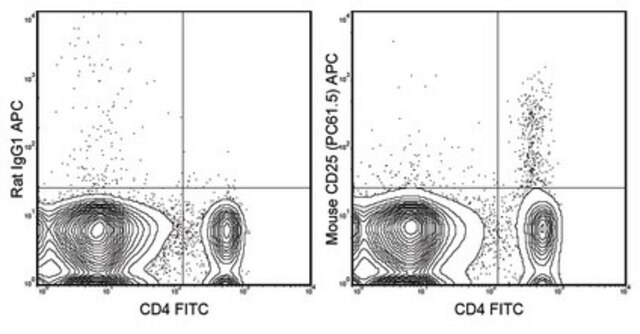
Flow Cytometry Analysis: A representative lot (pre-conjugated with Alexa Fluor 488) detected CLEC-2/CLEC1B expression on the surface of platelets and CD41-positive microparticles in platelet-rich plasma (PRP), but not on monocytes, neutrophils, dendritic cells, B- or T-cells (Gitz, E., et al. (2014). Blood. 124(14):2262-2270).

Function Assay: Clone AYP1 Fab fragment (2.5 µg/mL) cross-linked with anti-mouse Fab-specific F(ab)2 fragments, but not AYP1 Fab or anti-mouse F(ab)2 alone, induced surface P-selectin expression and the aggregation of washed human platelets (Gitz, E., et al. (2014). Blood. 124(14):2262-2270).

Immunoprecipitation Analysis: A representative lot immunoprecipitated CLEC-2/CLEC1B from human platelet lysates. Rhodocytin stimulation prior to cell lysis and IP induced CLEC-2/CLEC1B tyrosine phosphorylation (Gitz, E., et al. (2014). Blood. 124(14):2262-2270).
This mouse monoclonal Anti-CLEC-2 Antibody, clone AYP1, Cat. No. MABF957 is validated for use in Flow Cytometry, Function Assay, and Immunoprecipitation for the detection of CLEC-2.

Quality

Evaluated by Flow Cytometry in human platelets.

Flow Cytometry Analysis: 0.2 µL of this antibody detected CLEC-2/CLEC1B on the surface of human platelets.

Target description

26.60 kDa (isoform 1) and 23.10 kDa (isoform 2). ~32/40 kDa doublet reported due to differential glycosylation (Gitz, E., et al. (2014). Blood. 124(14):2262-2270).

Physical form

Format: Purified

Other Notes

Concentration: Please refer to lot specific datasheet.

Legal Information

ALEXA FLUOR is a trademark of Life Technologies

Not finding the right product?  

Try our Herramienta de selección de productos.

Storage Class

12 - Non Combustible Liquids

wgk_germany

WGK 2

flash_point_f

Not applicable

flash_point_c

Not applicable


Certificados de análisis (COA)

Busque Certificados de análisis (COA) introduciendo el número de lote del producto. Los números de lote se encuentran en la etiqueta del producto después de las palabras «Lot» o «Batch»

¿Ya tiene este producto?

Encuentre la documentación para los productos que ha comprado recientemente en la Biblioteca de documentos.

Visite la Librería de documentos

Nuestro equipo de científicos tiene experiencia en todas las áreas de investigación: Ciencias de la vida, Ciencia de los materiales, Síntesis química, Cromatografía, Analítica y muchas otras.

Póngase en contacto con el Servicio técnico