Saltar al contenido
MilliporeSigma
Todas las fotos(3)

Key Documents

MABC147

Sigma-Aldrich

Anti-TRAIL/CD253 Antibody, clone 6D12.2

clone 6D12.2, from mouse

Sinónimos:

Tumor necrosis factor ligand superfamily member 10, Apo-2 ligand, Apo-2L, TNF-related apoptosis-inducing ligand, Protein TRAIL, CD253

Iniciar sesiónpara Ver la Fijación de precios por contrato y de la organización


About This Item

UNSPSC Code:
12352203
eCl@ss:
32160702
NACRES:
NA.41

biological source

mouse

Quality Level

antibody form

purified immunoglobulin

antibody product type

primary antibodies

clone

6D12.2, monoclonal

species reactivity

human

technique(s)

flow cytometry: suitable
immunohistochemistry: suitable

isotype

IgG1κ

NCBI accession no.

UniProt accession no.

shipped in

wet ice

target post-translational modification

unmodified

Gene Information

human ... TNFSF10(8743)

General description

Apoptosis, or programmed cell death, occurs during normal cellular differentiation and development of multicellular organisms. Apoptosis is induced by certain cytokines including TNF and Fas ligand of the TNF family through their death domain containing receptors, TNFR1 and Fas. A novel member in the TNF family was recently identified and designated TRAIL (for TNF-related apoptosis-inducing ligand) and Apo-2L (for Apo-2 ligand). TRAIL is a type II membrane protein and is expressed in a variety of human tissues. Two novel death domain containing receptors DR4 and DR5 have been identified as the receptor for TRAIL. Like TNF and Fas ligand, TRAIL induces apoptosis and NF-kB activation in many tissues and cells.

Immunogen

GST-tagged recombinant protein corresponding to human TRAIL/CD253.

Application

Anti-TRAIL/CD253 Antibody, clone 6D12.2 is an antibody against TRAIL/CD253 for use in flow cytometry & IHC.
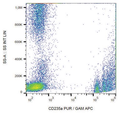
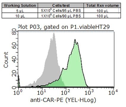
Flow Cytometry Analysis: 4 µg of this antibody detected TRAIL/CD253 in 1X10E6 human PBMC, which were treated with a proinflammatory stimulus.

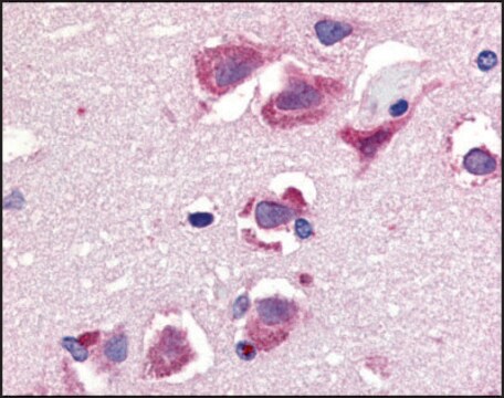
Immunohistochemistry Analysis: A 1:50 dilution from a representative lot detected TRAIL/CD253 in human prostate cancer sections. (Note: This antibody detects TRAIL/CD253 in cancer tissues and not in normal tissues.).
Research Category
Apoptosis & Cancer
Research Sub Category
Apoptosis - Additional

Quality

Evaluated by Flow Cytometry in HeLa cells, which were treated with a proinflammatory stimulus.

Flow Cytometry Analysis: 4 µg of this antibody detected TRAIL/CD253 in 1X10E6 HeLa cells, which were treated with a proinflammatory stimulus.

Target description

33 kDa calculated

Physical form

Format: Purified
Protein G Purified
Purified mouse monoclonal IgG1κ in buffer containing 0.1 M Tris-Glycine (pH 7.4), 150 mM NaCl with 0.05% sodium azide.

Storage and Stability

Stable for 1 year at 2-8°C from date of receipt.

Other Notes

Concentration: Please refer to the Certificate of Analysis for the lot-specific concentration.

Disclaimer

Unless otherwise stated in our catalog or other company documentation accompanying the product(s), our products are intended for research use only and are not to be used for any other purpose, which includes but is not limited to, unauthorized commercial uses, in vitro diagnostic uses, ex vivo or in vivo therapeutic uses or any type of consumption or application to humans or animals.

¿No encuentra el producto adecuado?  

Pruebe nuestro Herramienta de selección de productos.

Storage Class

12 - Non Combustible Liquids

wgk_germany

WGK 1

flash_point_f

Not applicable

flash_point_c

Not applicable


Certificados de análisis (COA)

Busque Certificados de análisis (COA) introduciendo el número de lote del producto. Los números de lote se encuentran en la etiqueta del producto después de las palabras «Lot» o «Batch»

¿Ya tiene este producto?

Encuentre la documentación para los productos que ha comprado recientemente en la Biblioteca de documentos.

Visite la Librería de documentos

Nuestro equipo de científicos tiene experiencia en todas las áreas de investigación: Ciencias de la vida, Ciencia de los materiales, Síntesis química, Cromatografía, Analítica y muchas otras.

Póngase en contacto con el Servicio técnico