17-441
Rac1/Cdc42 Activation Assay Kit
The Rac1/Cdc42 Activation Assay provides an effective method for detecting Rac & Cdc42 activity in cell lysates.
Sinónimos:
Rac1 activation assay
Iniciar sesiónpara Ver la Fijación de precios por contrato y de la organización
About This Item
UNSPSC Code:
12161503
eCl@ss:
32161000
NACRES:
NA.84
Productos recomendados
Quality Level
species reactivity
human, mouse, rat
manufacturer/tradename
Upstate®
technique(s)
activity assay: suitable
affinity binding assay: suitable (G-protein)
NCBI accession no.
UniProt accession no.
shipped in
wet ice
Gene Information
human ... RAC1(5879)
General description
The Rac1/Cdc42 Activation Assay (Cat. No. 17-441) provides an effective method for detecting Rac and Cdc42 activity in cell lysates. This assay uses the downstream effector of Rac/Cdc42, p21-activated protein kinase (PAK1), to isolated the active GTP-bound form of Rac/Cdc42 from the sample. The p21 binding domain (PBD) of PAK1 is expressed as a GST-fusion protein and coupled to agarose beads. After precipitation, an immunoblot is performed and the activated Rac/Cdc42 is detected with specific monoclonal antibodies, followed by HRP-conjugated secondary antibody and ECL reagent.
Specificity
Species Cross-reactivity: Human and mouse. Predicted to cross-react with all mammalian species.
Anti-Rac1, clone 23A8: Species Cross-reactivity: Human, mouse and rat. Other species cross-reactivity unknown.
Anti-cdc42 Species Cross-reactivity: Human, mouse, rat and dog. Other species cross-reactivity unknown.
Anti-Rac1, clone 23A8: Species Cross-reactivity: Human, mouse and rat. Other species cross-reactivity unknown.
Anti-cdc42 Species Cross-reactivity: Human, mouse, rat and dog. Other species cross-reactivity unknown.
Components
Rac/cdc42 Assay Reagent (PAK-1 PBD, agarose), Catalog # 14-325 :One vial containing 300 μg of PAK-1 PBD bound to 150 μL of glutathione agarose beads, provided as a 50% slurry in 20mM PBS, pH 7.4, containing 50% glycerol for a final volume of 300 μL.
Anti-Rac1, clone 23A8, Catalog #05-389: One vial containing 250 μg of protein G purified mouse IgG2b in 250 μL of storage buffer (0.1 M Tris-glycine, pH 7.4, 0.15 M NaCl, containing 0.05% sodium azide).
Anti-cdc42, (mouse monoclonal IgG1) , Catalog # 05-542: One vial containing 50 μg of purified mouse IgG1 in 200 μL of 50% storage buffer (20 mM sodium phosphate, pH 7.5, 150 mM NaCl, 1.5 mM sodium azide containing, 1 mg/mL BSA) and 50% glycerol.
Mg2+ Lysis/Wash Buffer, 5X, Catalog # 20-168 : Two vials, each vial containing 18 mL of 5X MLB: 125 mM HEPES, pH 7.5, 750 mM NaCl, 5% Igepal CA-630, 50 mM MgCl2, 5 mM EDTA and 10% glycerol.
100X GTPγS, 10mM, Catalog # 20-176: One vial containing 50 μL of 10 mM GTPγS, 100X stock, in 50 mM Tris-HCl, pH 7.8, non-hydrolyzable analog of GTP. Sufficient to label 5 mL of cell lysates.
100X GDP, 100mM, Catalog # 20-177: One vial containing 50 μL of 100 mM GDP, 100X stock, in 50 mM Tris-HCl, pH 7.8. GDP (Guanosine 5′-Diphosphate) for in vitro labeling of G-proteins in the inactive form. Sufficient to label 5ml of cell lysates.
Quality
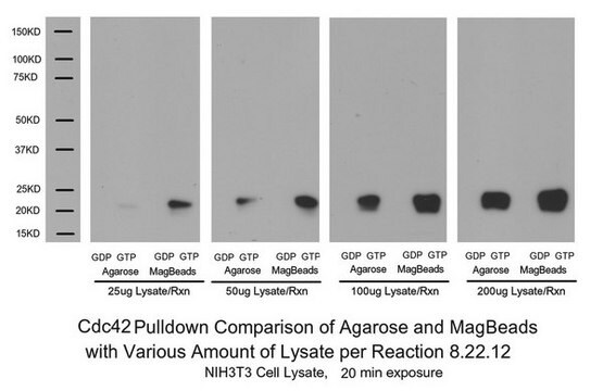
Routinely evaluated by precipitating Rac1-GTP and cdc42-GTP from 3T3/A31 and HeLa cell lysates that had been loaded with GTPγS(lysates were loaded in vitro with GTPγS for cdc42-GTP). The precipitated Rac1-GTP and cdc42-GTP was detected by immunoblot analysis using anti-Rac1 (1 μg/ml).
Storage and Stability
1 year at -20°C from date of shipment
Legal Information
UPSTATE is a registered trademark of Merck KGaA, Darmstadt, Germany
Disclaimer
Unless otherwise stated in our catalog or other company documentation accompanying the product(s), our products are intended for research use only and are not to be used for any other purpose, which includes but is not limited to, unauthorized commercial uses, in vitro diagnostic uses, ex vivo or in vivo therapeutic uses or any type of consumption or application to humans or animals.
signalword
Danger
hcodes
Hazard Classifications
Aquatic Acute 1 - Aquatic Chronic 2 - Eye Dam. 1
Storage Class
10 - Combustible liquids
Certificados de análisis (COA)
Busque Certificados de análisis (COA) introduciendo el número de lote del producto. Los números de lote se encuentran en la etiqueta del producto después de las palabras «Lot» o «Batch»
¿Ya tiene este producto?
Encuentre la documentación para los productos que ha comprado recientemente en la Biblioteca de documentos.
Krishna Chinthalapudi et al.
Nature communications, 9(1), 1338-1338 (2018-04-08)
Neurofibromatosis type 2 (NF2) is a tumor-forming disease of the nervous system caused by deletion or by loss-of-function mutations in NF2, encoding the tumor suppressing protein neurofibromin 2 (also known as schwannomin or merlin). Neurofibromin 2 is a member of
Marco Trerotola et al.
Cancer research, 73(10), 3155-3167 (2013-03-29)
The molecular mechanisms underlying metastatic dissemination are still not completely understood. We have recently shown that β(1) integrin-dependent cell adhesion to fibronectin and signaling is affected by a transmembrane molecule, Trop-2, which is frequently upregulated in human carcinomas. Here, we
Mitsuyoshi Motizuki et al.
The Journal of biological chemistry, 296, 100545-100545 (2021-03-21)
Transforming growth factor-β (TGF-β) signaling promotes cancer progression. In particular, the epithelial-mesenchymal transition (EMT) induced by TGF-β is considered crucial to the malignant phenotype of cancer cells. Here, we report that the EMT-associated cellular responses induced by TGF-β are mediated
Enikő Tátrai et al.
Oncotarget, 8(27), 44498-44510 (2017-06-01)
Tumor hypoxia promotes neoangiogenesis and contributes to the radio- and chemotherapy resistant and aggressive phenotype of cancer cells. However, the migratory response of tumor cells and the role of small GTPases regulating the organization of cytoskeleton under hypoxic conditions have
Tao Dong et al.
Proceedings of the National Academy of Sciences of the United States of America, 113(27), 7644-7649 (2016-06-24)
The etiology of autism is so complicated because it involves the effects of variants of several hundred risk genes along with the contribution of environmental factors. Therefore, it has been challenging to identify the causal paths that lead to the
Nuestro equipo de científicos tiene experiencia en todas las áreas de investigación: Ciencias de la vida, Ciencia de los materiales, Síntesis química, Cromatografía, Analítica y muchas otras.
Póngase en contacto con el Servicio técnico
