Saltar al contenido
MilliporeSigma
Todas las fotos(3)

Documentos clave

06-735

Sigma-Aldrich

Anti-Caspase 3 Antibody

Upstate®, from rabbit

Sinónimos:

Anti-CASP-3, Anti-Caspase-3

Iniciar sesiónpara Ver la Fijación de precios por contrato y de la organización


About This Item

UNSPSC Code:
12352203
eCl@ss:
32160702
NACRES:
NA.41

biological source

rabbit

Quality Level

antibody form

purified immunoglobulin

antibody product type

primary antibodies

clone

polyclonal

species reactivity

mouse, human, rat

manufacturer/tradename

Upstate®

technique(s)

immunohistochemistry: suitable
western blot: suitable

isotype

IgG

NCBI accession no.

UniProt accession no.

shipped in

wet ice

target post-translational modification

unmodified

Gene Information

human ... CASP3(836)

General description

Caspase-3 (UniProt: P42574; also known as EC:3.4.22.56, CASP-3, Apopain, Cysteine protease CPP32, CPP-32, Protein Yama, SREBP cleavage activity 1, SCA-1) is encoded by the CASP3 (also known as CPP32) gene (Gene ID: 836) in human. Cysteine-aspartic proteases or Caspases play essential roles in apoptosis, necrosis, and inflammation. Historically, caspases were numbered in the order in which they were identified. Caspase-3 is a heterotetrameric enzyme that consists of two anti-parallel arranged heterodimers, each one formed by a 17 kDa (p17) and a 12 kDa (p12) subunit. Caspase-3 is initially produced with a propeptide sequence (aa 1-9), the removal of which yields the 268 aa. caspase-3 proenzyme. Upon activation, the proenzyme is proteolytically cleaved first between Asp175-Ser176 to generate a p20 (aa 10-175) fragment and the p12 (aa 176-277) subunit. Further cleavage of the p20 fragment between Asp28-Ser29 produces the p17 (aa 29-175) subunit. The p17 and p12 subunits dimerize and forms the active caspase-3 enzyme. Caspase-3 has a strict requirement for an Asp residue at positions P1 and P4. It has a preferred cleavage sequence of Asp-Xaa-Xaa-Asp-|- with a hydrophobic amino-acid residue at P2 and a hydrophilic amino-acid residue at P3, although Val or Ala are also accepted at this position. Caspase-3 is involved in the activation cascade of caspases responsible for apoptosis execution. At the onset of apoptosis, it proteolytically cleaves poly(ADP-ribose) polymerase (PARP) at a Asp216-|-Gly217 bond. Caspase-3 mediates the proteolytic activation of caspases-6 and -7, while caspase-3 itself is processed and activated by caspase-8, -9, or -10.

Specificity

Recognizes full-length Caspase 3 (Yama/Apopain) and proteolytic fragments.

Immunogen

Human full-length Caspase 3 fusion protein containing a histidine-6 tag

Application

This Anti-Caspase 3 Antibody is validated for use in Immunihistochmistry and Western Blotting for the detection of Caspase 3.
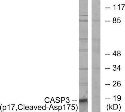
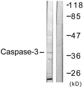
Western Blotting Analysis: 1μg/mL of this antibody detects Caspase-3 in A431 cell lysate.

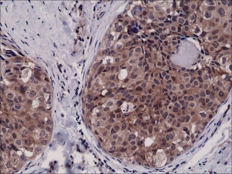
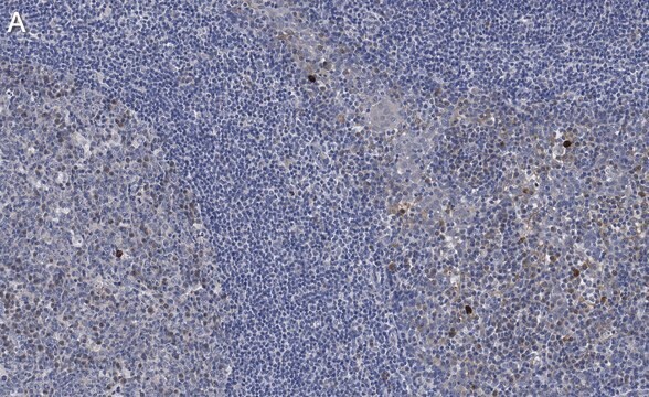
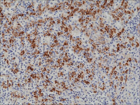
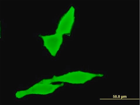
Immunohistochemistry (Paraffin) Analysis: A 1:250 dilution of this antibody detected Caspase-3 in Human tonsil tissue sections.

Quality

routinely evaluated by immunoblot on RIPA lysates from non-stimulated human A431 cells, mouse 3T3/A31 or rat PC12 cells

Target description

32 kDa

Linkage

Replaces: 04-1090; 04-439

Physical form

Format: Purified
Protein A purified IgG in of 0.1M Tris-glycine, pH 7.4, 0.15M NaCl,and 0.05% sodium azide.

Storage and Stability

Stable for 2 years at 2-8°C from date of shipment. For maximum recovery of product, centrifuge the original vial prior to removing the cap.

Analysis Note

Control
Positive Antigen Control: Catalog #12-301, non-stimulated A431 cell lysate. Add 2.5µL of 2-mercaptoethanol/100µL of lysate and boil for 5 minutes to reduce the preparation. Load 20µg of reduced lysate per lane for mingels.

Legal Information

UPSTATE is a registered trademark of Merck KGaA, Darmstadt, Germany

¿No encuentra el producto adecuado?  

Pruebe nuestro Herramienta de selección de productos.

Optional

Storage Class

10 - Combustible liquids

wgk_germany

WGK 1


Certificados de análisis (COA)

Busque Certificados de análisis (COA) introduciendo el número de lote del producto. Los números de lote se encuentran en la etiqueta del producto después de las palabras «Lot» o «Batch»

¿Ya tiene este producto?

Encuentre la documentación para los productos que ha comprado recientemente en la Biblioteca de documentos.

Visite la Librería de documentos

Role of p53 and ATM in photodynamic therapy-induced apoptosis.
Viola Heinzelmann-Schwarz, Andre Fedier, Rene Hornung, Heinrich Walt, Urs Haller, Daniel Fink
Lasers in Surgery and Medicine null
M Leist et al.
Biochemical and biophysical research communications, 258(1), 215-221 (1999-05-01)
The endogenous mediator nitric oxide (NO) blocked apoptosis of Jurkat cells elicited by staurosporine, anti-CD95 or chemotherapeutics, and switched death to necrosis. The switch in the mode of cell death was dependent on the ATP loss elicited by NO. This
A caspase cascade regulating developmental axon degeneration.
Simon, DJ; Weimer, RM; McLaughlin, T; Kallop, D; Stanger, K; Yang, J; O'Leary et al.
The Journal of Neuroscience null
Oral inoculation of probiotics Lactobacillus acidophilus NCFM suppresses tumour growth both in segmental orthotopic colon cancer and extra-intestinal tissue.
Chen, CC; Lin, WC; Kong, MS; Shi, HN; Walker, WA; Lin, CY; Huang, CT; Lin, YC; Jung, SM; Lin, TY
The British Journal of Nutrition null
Wafaa S Ramadan et al.
Cells, 10(12) (2021-12-25)
Patients suffering from Alzheimer's disease (AD) are still increasing worldwide. The development of (AD) is related to oxidative stress and genetic factors. This study investigated the therapeutic effects of ellagic acid (EA) on the entorhinal cortex (ERC), which plays a

Nuestro equipo de científicos tiene experiencia en todas las áreas de investigación: Ciencias de la vida, Ciencia de los materiales, Síntesis química, Cromatografía, Analítica y muchas otras.

Póngase en contacto con el Servicio técnico