03-206
RIPAb+ hnRNP U - RIP Validated Antibody and Primer Set
clone 3G6, from mouse
Sinónimos:
Scaffold attachment factor A, heterogeneous nuclear ribonucleoprotein U, heterogeneous nuclear ribonucleoprotein U (scaffold attachment factor A), hnRNP U, hnRNP U protein, p120 nuclear protein
About This Item
Productos recomendados
biological source
mouse
Quality Level
antibody form
purified immunoglobulin
clone
3G6, monoclonal
species reactivity
human
manufacturer/tradename
RIPAb+
Upstate®
technique(s)
RIP: suitable
western blot: suitable
isotype
IgG2bκ
NCBI accession no.
UniProt accession no.
shipped in
dry ice
General description
Proteins of the heterogeneous nuclear ribonucleoparticles (hnRNP) family form a structurally diverse group of RNA binding proteins implicated in various functions. Recently, hnRNP proteins have been shown to hinder communication between factors bound to different splice sites. hnRNP-U, also termed scaffold attachment factor A (SAF-A), binds to pre-mRNA and nuclear matrix/scaffold attachment region DNA elements.
Specificity
Immunogen
Application
Representative lot data.
RIP lysate from HeLa cells (~2 X 10E7 cell equivalents per IP) was subjected to immunoprecipitation using 5 µg of either a normal mouse IgG, (Cat. # CS200621), or 5 µg of Anti-hnRNP U antibody (Cat. # CS207320). ten percent of the precipitated proteins (lane 1: normal mouse IgG, lane 2: hnRNP U) and HeLa whole cell lysate (lane 3) were resolved by electrophoresis, transferred to nitrocellulose and probed with anti-hnRNP U antibody (Cat. # CS207320, 1:1000). Proteins were visualized using One-Step IP-Western kit (GenScript Cat. # L00231).
Arrow indicates hnRNP U. (Figure 2).
Automated Microfluidics-based Electrophoretic RNA Separation and Analysis (MFE):
Representative lot data.
RIP Lysate prepared from HeLa cells (2 X 10E7 cell equivalents per IP) were subjected to immunoprecipitation using 5 µg of either 1. normal mouse IgG (Cat. # CS200621), or 2. Anti-hnRNP U antibody (Cat. # CS207320) and the Magna RIP RNA-Binding Protein Immunoprecipitation Kit (Cat. # 17-700).
Successful immunoprecipitation of hnRNP U-associated RNA was verified by automated microfluidics-based electrophoretic RNA separation and analysis. Please refer to the Magna RIP (Cat. # 17-700) or EZ-Magna RIP (Cat. # 17-701) protocol for experimental details. Electropherograms were generated by plotting fluorescence intensities versus migration times (Figure 3A). The virtual gel view was created from this data (Figure 3B).
Western Blot Analysis:
Representative lot data.
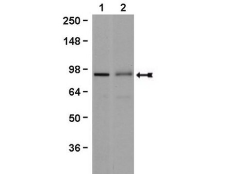
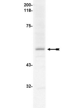
K562 cell lysate was probed with Anti-hnRNP U, clone 3G6 (0.01 µg/mL). Proteins were visualized using a Goat Anti-Mouse IgG secondary antibody conjugated to HRP and a chemiluminescence detection system.
Arrow indicates hnRNP U (~120 kDa). (Figure 4).
Epigenetics & Nuclear Function
RNA Metabolism & Binding Proteins
Apoptosis - Additional
Packaging
Quality
Representative lot data.
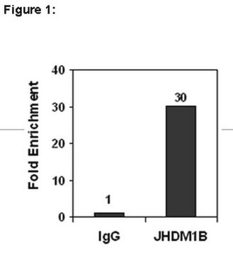
RIP Lysate prepared from HeLa cells (2 X 10E7 cell equivalents per IP) were subjected to immunoprecipitation using 5 µg of either a normal mouse IgG or 5 µg of Anti-hnRNP U antibody and the Magna RIP® RNA-Binding Protein Immunoprecipitation Kit (Cat. # 17-700).
Successful immunoprecipitation of hnRNP U-associated RNA was verified by qPCR using RIP Primers Ribosomal Protein S19, (Figure 1).
Please refer to the Magna RIP (Cat. # 17-700) or EZ-Magna RIP (Cat. # 17-701) protocol for experimental details.
Target description
Physical form
Normal Mouse IgG, Part # CS200621. One vial containing 125 µg of purified mouse IgG in 125 µL of storage buffer containing 0.1% sodium azide. Store at -20°C.
RIP Primers, Ribosomal Protein S19, Part # CS207321. One vial containing 75 μL of 5 μM of each primer specific for human c-myc 3′ UTR. Store at -20°C.
FOR: ACG CGA GCT GCT TCC ACA G
REV: AGC TGC CAC CTG TCC GGC
Storage and Stability
Handling Recommendations: Upon receipt, and prior to removing the cap, centrifuge the vial and gently mix the solution. Aliquot into microcentrifuge tubes and store at -20°C. Avoid repeated freeze/thaw cycles, which may damage IgG and affect product performance. Note: Variabillity in freezer temperatures below -20°C may cause glycerol containing solutions to become frozen during storage.
Analysis Note
Includes negative control normal mouse IgG antibody and control primers specific for the cDNA of human Ribosomal Protein S19.
Other Notes
Legal Information
Disclaimer
Storage Class
10 - Combustible liquids
Certificados de análisis (COA)
Busque Certificados de análisis (COA) introduciendo el número de lote del producto. Los números de lote se encuentran en la etiqueta del producto después de las palabras «Lot» o «Batch»
¿Ya tiene este producto?
Encuentre la documentación para los productos que ha comprado recientemente en la Biblioteca de documentos.
Nuestro equipo de científicos tiene experiencia en todas las áreas de investigación: Ciencias de la vida, Ciencia de los materiales, Síntesis química, Cromatografía, Analítica y muchas otras.
Póngase en contacto con el Servicio técnico
