推薦產品
形狀
powder or crystals
品質等級
成份
Dye content, 50%
mp
>300 °C
λmax
229 nm (2nd)
340-355 nm
應用
diagnostic assay manufacturing
hematology
histology
儲存溫度
room temp
SMILES 字串
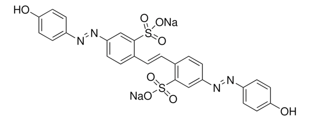
[Na+].Cc1ccc2nc(sc2c1S([O-])(=O)=O)-c3ccc4nc(sc4c3)-c5ccc(N)cc5
InChI
1S/C21H15N3O3S3.Na/c1-11-2-8-16-18(19(11)30(25,26)27)29-21(24-16)13-5-9-15-17(10-13)28-20(23-15)12-3-6-14(22)7-4-12;/h2-10H,22H2,1H3,(H,25,26,27);/q;+1/p-1
InChI 密鑰
RSRNHSYYBLEMOI-UHFFFAOYSA-M
尋找類似的產品? 前往 產品比較指南
應用
Primuline 是制备叶绿体脂质膜脂质的一种试剂。它被用作制备薄层色谱(TLC)中的染料。
生化/生理作用
樱草黄,也称为直接黄 59 或樱草灵,是一种更敏感和非破坏性的脂质染料。它广泛用作制备型薄层色谱(TLC)中的荧光染料。樱草黄适用于活体染色。它用作植物组织的荧光染料。樱草黄还用于染色花粉粒和木质化细胞壁。此外,它还有助于分离完整和破碎的淀粉粒。在 pH8 时,樱草黄用作肥大细胞的荧光染料。
儲存類別代碼
11 - Combustible Solids
水污染物質分類(WGK)
WGK 3
閃點(°F)
Not applicable
閃點(°C)
Not applicable
個人防護裝備
Eyeshields, Gloves, type N95 (US)
客戶也查看了
T Taki et al.
Analytical biochemistry, 223(2), 232-238 (1994-12-01)
A new and simple method for purifying glycosphingolipids and phospholipids by using "TLC blotting" was established. Glycosphingolipids separated by two-dimensional thin-layer chromatography (TLC) were made visible with primuline reagent, and then bands were marked with a drawing colored pencil. The
[New data on the organization of descending hypothalamic pathways obtained by the method of double retrograde labeling of neurons with fluorochromes].
N Z Doroshenko et al.
Doklady Akademii nauk SSSR, 282(1), 232-236 (1985-05-01)
J H Duffus et al.
Stain technology, 59(2), 79-82 (1984-03-01)
Treatment of cells and purified cell walls of the fission yeast Schizosaccharomyces pombe with primuline reveals the septum as a bright fluorescent band. When polysaccharides containing (1----3)-beta-, (1----6)-beta- or (1----3)-alpha-glucosidic linkages are treated with primuline, only those molecules containing chains
V A Maisky et al.
Journal of the autonomic nervous system, 34(2-3), 119-128 (1991-06-15)
The organization of the neuroanatomical substrate which provides the supraspinal catecholaminergic innervation of the upper thoracic spinal cord in the rat was studied by means of retrograde labelling of neurons with primuline and other dyes, combined with simultaneous catecholamine fluorescence
K Imamoto
Archivum histologicum Japonicum = Nihon soshikigaku kiroku, 47(3), 271-277 (1984-08-01)
Scanning electron microscopy revealed numerous macrophagic ameboid cells on the ependymal surface of all brain ventricles in neonatal rats. Macrophagic ameboid cells aggregated in the sulcus medianus of the fossa rhomboidea, the recessus of the cerebral aqueduct and the recessus
我們的科學家團隊在所有研究領域都有豐富的經驗,包括生命科學、材料科學、化學合成、色譜、分析等.
聯絡技術服務