推薦產品
生物源
rat
品質等級
抗體表格
purified immunoglobulin
抗體產品種類
primary antibodies
無性繁殖
TROMA-3, monoclonal
物種活性
mouse, human
包裝
antibody small pack of 25 μg
技術
electron microscopy: suitable
immunofluorescence: suitable
immunohistochemistry: suitable (paraffin)
immunoprecipitation (IP): suitable
western blot: suitable
同型
IgG2aκ
NCBI登錄號
UniProt登錄號
運輸包裝
ambient
目標翻譯後修改
unmodified
基因資訊
human ... KRT19(3880)
mouse ... Krt19(16669)
一般說明
Keratin, type I cytoskeletal 19 (UniProt: P19001; also known as Cytokeratin-19, CK-19, Keratin-19, K19) is encoded by the Krt19 (also known as Krt1-19) gene (Gene ID: 16669) in murine species. CK-19 belongs to the family of intermediate filament (IF) family, but differs from other IF proteins in lacking the C-terminal tail domain. About 20 different cytokeratin proteins have been reported in higher animals in varying combinations during various stages of development. CK-19 is normally expressed in the lining of the gastroenteropancreatic and hepatobiliary tracts. It is expressed throughout embryonic development with highest levels observed at 8.5 days post coitum. Its expression is shown to decrease by day 11.5 and then is increased again by 17.5 days post coitum. CK-19 is shown to be involved in the organization of myofibrils and together with keratin 8, it helps to link the contractile apparatus to dystrophin at the costameres of striated muscle. CK-19 has been shown to be an independent prognostic factor for pancreatic neuroendocrine tumors, especially the insulin-negative tumors. TROMA 3 has been found to stain trophoblastoma TDM.1 cells, a subpopulation of the retinoic acid.-induced F9 and PCC3/ND1 cells, and in 5-azacytidine-treated 1246 cells.
特異性
Clone TROMA-3 detects cytokeratin 19 in human and murine cells.
免疫原
Purified intermediate filaments from DM1 (Trophoblatoma) cell line.
應用
Anti-Cytokeratin 19, clone TROMA-3, Cat. No. MABT913, is a rat monoclonal antibody that detects Keratin, type I cytoskeletal 19 and is tested for use in Electron Microscopy, Immunofluorescence, Immunohistochemistry (Paraffin), Immunoprecipitation, and Western Blotting.
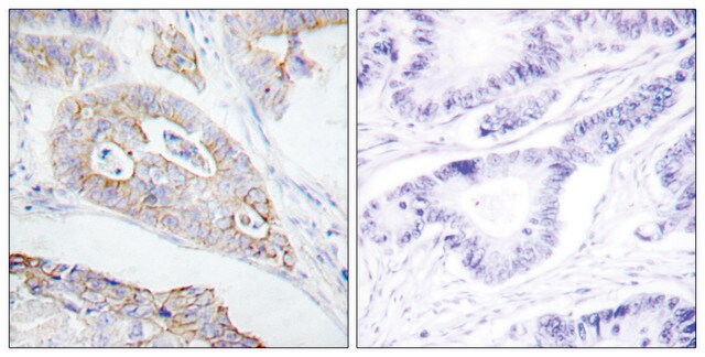
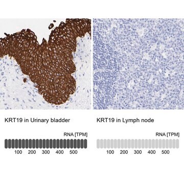
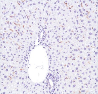
Immunohistochemistry Analysis: A 1:250 dilution from a representative lot detected Cytokeratin 19 in mouse lung tissue.
Western Blotting Analysis: A representative lot detected Cytokeratin 19 in Western Blotting applications (Boller, K., et. al. (1987). Eur J Cell Biol. 43(3):459-68).
Electron Microscopy Analysis: A representative lot detected Cytokeratin 19 in Electron Microscopy applications (Boller, K., et. al. (1987). Eur J Cell Biol. 43(3):459-68).
Immunofluorescence Analysis: A representative lot detected Cytokeratin 19 in Immunofluorescence applications (Boller, K., et. al. (1987). Eur J Cell Biol. 43(3):459-68).
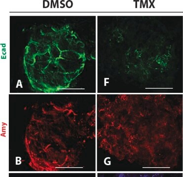
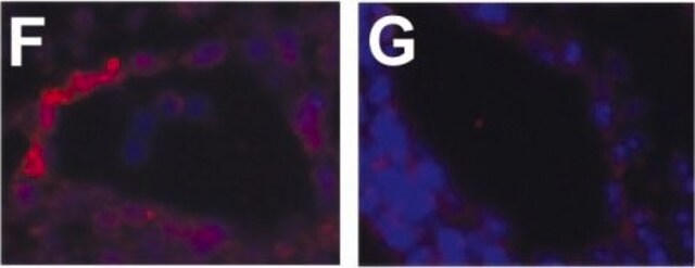
Immunohistochemistry Analysis: A representative lot detected Cytokeratin 19 in Immunohistochemistry applications (Takase, H.M., et. al. (2013). Genes Dev. 27(2):169-81).
Immunoprecipitation Analysis: A representative lot detected Cytokeratin 19 in Immunoprecipitation applications (Ichinose, Y., et. al. (1990). Biochem Biophys Res Commun. 167(2):644-7).
品質
Evaluated by Western Blotting in MCF-7 cell lysate.
Western Blotting Analysis: 0.5 µg/mL of this antibody detected Cytokeratin 19 in 10 µg of MCF-7 cell lysate.
Western Blotting Analysis: 0.5 µg/mL of this antibody detected Cytokeratin 19 in 10 µg of MCF-7 cell lysate.
標靶描述
~44 kDa observed; 44.54 kDa calculated. Uncharacterized bands may be observed in some lysate(s).
外觀
Format: Purified
其他說明
Concentration: Please refer to lot specific datasheet.
未找到適合的產品?
試用我們的產品選擇工具.
儲存類別代碼
12 - Non Combustible Liquids
水污染物質分類(WGK)
WGK 1
閃點(°F)
Not applicable
閃點(°C)
Not applicable
分析證明 (COA)
輸入產品批次/批號來搜索 分析證明 (COA)。在產品’s標籤上找到批次和批號,寫有 ‘Lot’或‘Batch’.。
Ana Caroline Costa-da-Silva et al.
iScience, 25(1), 103592-103592 (2022-01-11)
Chronic graft-versus-host disease (cGVHD) targets include the oral mucosa and salivary glands after allogeneic hematopoietic stem cell transplant (HSCT). Without incisional biopsy, no diagnostic test exists to confirm oral cGVHD. Consequently, therapy is often withheld until severe manifestations develop. This
Rebecca Marcus et al.
Cancers, 13(22) (2021-11-28)
Intrahepatic cholangiocarcinoma (ICC) is a primary biliary malignancy that harbors a dismal prognosis. Oncogenic mutations of KRAS and loss-of-function mutations of BRCA1-associated protein 1 (BAP1) have been identified as recurrent somatic alterations in ICC. However, an autochthonous genetically engineered mouse
Romina Lasagni Vitar et al.
Stem cell reports, 17(4), 849-863 (2022-03-26)
Severe ocular surface diseases can lead to limbal stem cell deficiency (LSCD), which is accompanied by defective healing. We aimed to evaluate the role of the substance P (SP)/neurokinin-1 receptor (NK1R) pathway in corneal epithelium wound healing in a pre-clinical
我們的科學家團隊在所有研究領域都有豐富的經驗,包括生命科學、材料科學、化學合成、色譜、分析等.
聯絡技術服務
