推薦產品
生物源
mouse
品質等級
抗體表格
purified immunoglobulin
抗體產品種類
primary antibodies
無性繁殖
BV6, monoclonal
物種活性
human
技術
flow cytometry: suitable
immunocytochemistry: suitable
immunohistochemistry: suitable (paraffin)
western blot: suitable
同型
IgG2aκ
NCBI登錄號
UniProt登錄號
運輸包裝
wet ice
目標翻譯後修改
unmodified
基因資訊
human ... CDH5(1003)
一般說明
人血管内皮 (VE)-钙粘蛋白是一种严格位于细胞与细胞连接处的钙依赖性粘附分子。VE-钙黏着蛋白存在于所有类型的内皮(静脉、动脉、毛细血管和大血管)中。钙黏着蛋白是钙依赖性细胞粘附蛋白。它们优先在连接细胞中以同质方式与自身相互作用。因此,钙黏着蛋白可能有助于异质细胞类型的分类。VE-钙粘蛋白可能通过控制细胞间连接的凝聚力和组织在内皮细胞生物学中起重要作用。VE-钙粘蛋白还与α连环蛋白结合形成与细胞骨架的连接。
免疫原
HUVEC细胞
應用
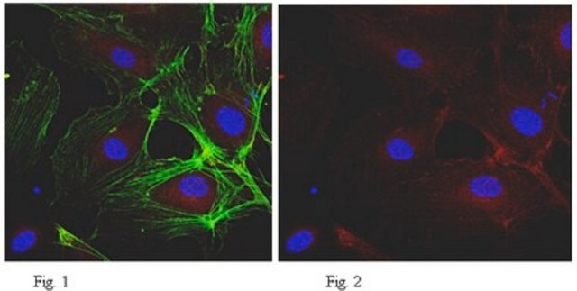
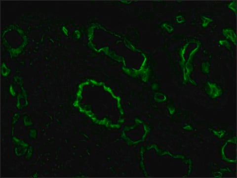
免疫细胞化学分析:代表性批次的 1:500 稀释液在 HUVEC 细胞的细胞-细胞连接处检测到 VE-钙粘蛋白。
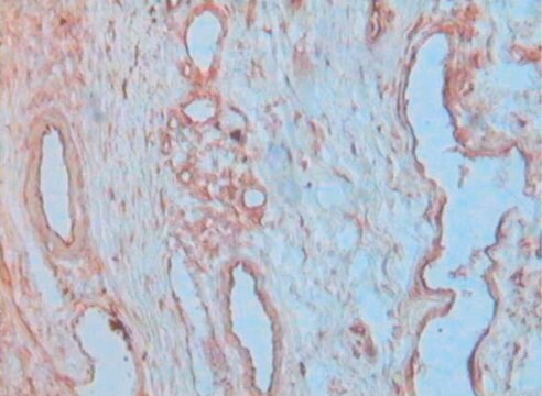
免疫组织化学分析:独立实验室将代表性批次的 1:10-1:20 稀释液用于石蜡包埋的组织切片。建议使用缓冲福尔马林固定,固定时间不超过 12 小时。还建议在柠檬酸盐缓冲液(货号 21545)中进行高温抗原修复。抗体还可用于使用免疫过氧化物酶染色方案标记丙酮固定的低温切片或细胞(例如 IHC Select 检测试剂盒,货号 DAB150)
流式细胞术分析:代表性批次在 Ca2+ 存在下使用。注意:使用 PBS + 2-5mM EDTA 进行细胞分离。
蛋白质印迹分析:可能需要非还原条件,缓冲液中需要 Ca2+ (2-5 mM)。
免疫组织化学分析:独立实验室将代表性批次的 1:10-1:20 稀释液用于石蜡包埋的组织切片。建议使用缓冲福尔马林固定,固定时间不超过 12 小时。还建议在柠檬酸盐缓冲液(货号 21545)中进行高温抗原修复。抗体还可用于使用免疫过氧化物酶染色方案标记丙酮固定的低温切片或细胞(例如 IHC Select 检测试剂盒,货号 DAB150)
流式细胞术分析:代表性批次在 Ca2+ 存在下使用。注意:使用 PBS + 2-5mM EDTA 进行细胞分离。
蛋白质印迹分析:可能需要非还原条件,缓冲液中需要 Ca2+ (2-5 mM)。
研究子类别
粘附(CAMs)
粘附(CAMs)
研究类别
细胞结构
细胞结构
该抗 VE-钙粘蛋白抗体克隆 BV6 经过验证,可用于 WB、IC、FC、IH(P) 中检测 VE-钙粘蛋白。
品質
通过蛋白质印迹对HUVEC细胞裂解物进行了评估。
蛋白质印迹分析:该抗体以 1:2,000 稀释在 HUVEC 细胞裂解物中检测到 VE-钙粘蛋白。
蛋白质印迹分析:该抗体以 1:2,000 稀释在 HUVEC 细胞裂解物中检测到 VE-钙粘蛋白。
標靶描述
观测值〜120 kDa。
该蛋白的计算分子量为 82 kDa,但由于该蛋白质被糖基化,因此其分子量将在 ~90-140 kDa 之间。
该蛋白的计算分子量为 82 kDa,但由于该蛋白质被糖基化,因此其分子量将在 ~90-140 kDa 之间。
聯結
替代:MAB1989
外觀
形式:纯化
纯化的小鼠单克隆IgG2aκ,溶于含有0.1 M Tris-甘氨酸(pH 7.4,150 mM NaCl)和0.05%叠氮化钠的缓冲液中。
蛋白G纯化
儲存和穩定性
自收到之日起,在2-8°C条件下可稳定保存1年。
分析報告
对照
HUVEC细胞裂解液
HUVEC细胞裂解液
其他說明
浓度:请参考批次特异性浓缩物的检验报告。
免責聲明
除非我们的产品目录或产品附带的其他公司文档另有说明,否则我们的产品仅供研究使用,不得用于任何其他目的,包括但不限于未经授权的商业用途、体外诊断用途、离体或体内治疗用途或任何类型的消费或应用于人类或动物。
未找到適合的產品?
試用我們的產品選擇工具.
儲存類別代碼
12 - Non Combustible Liquids
水污染物質分類(WGK)
WGK 1
閃點(°F)
Not applicable
閃點(°C)
Not applicable
分析證明 (COA)
輸入產品批次/批號來搜索 分析證明 (COA)。在產品’s標籤上找到批次和批號,寫有 ‘Lot’或‘Batch’.。
B Herren et al.
Molecular biology of the cell, 9(6), 1589-1601 (1998-06-17)
Growth factor deprivation of endothelial cells induces apoptosis, which is characterized by membrane blebbing, cell rounding, and subsequent loss of cell-matrix and cell-cell contacts. In this study, we show that initiation of endothelial apoptosis correlates with cleavage and disassembly of
Functional roles for PECAM-1 (CD31) and VE-cadherin (CD144) in tube assembly and lumen formation in three-dimensional collagen gels.
Yang, S; Graham, J; Kahn, JW; Schwartz, EA; Gerritsen, ME
The American Journal of Pathology null
M Harraz et al.
Stem cells (Dayton, Ohio), 19(4), 304-312 (2001-07-21)
A subset of adult peripheral blood leukocytes functions as endothelial cell progenitors called angioblasts. They can incorporate into the vasculature in animal models of neovascularization and accelerate the restoration of blood flow to mouse ischemic limbs. Earlier reports suggested that
Noritoshi Nagaya et al.
Circulation, 108(7), 889-895 (2003-07-02)
Circulating endothelial progenitor cells (EPCs) migrate to injured vascular endothelium and differentiate into mature endothelial cells. We investigated whether transplantation of vasodilator gene-transduced EPCs ameliorates monocrotaline (MCT)-induced pulmonary hypertension in rats. We obtained EPCs from cultured human umbilical cord blood
Pan Liu et al.
Biotechnology and bioengineering, 118(1), 423-432 (2020-09-25)
Vascular leak is a key driver of organ injury in diseases, and strategies that reduce enhanced permeability and vascular inflammation are promising therapeutic targets. Activation of the angiopoietin-1 (ANG1)-Tie2 tyrosine kinase signaling pathway is an important regulator of vascular quiescence.
我們的科學家團隊在所有研究領域都有豐富的經驗,包括生命科學、材料科學、化學合成、色譜、分析等.
聯絡技術服務