跳轉至內容
Merck
全部照片(2)

重要文件

AB2971

Sigma-Aldrich

抗LAMP-1(CD107a)抗体

from rabbit, purified by affinity chromatography

同義詞:

lysosomal-associated membrane protein 1, lysosome-associated membrane glycoprotein 1, Lysosome-associated membrane protein 1, CD107 antigen-like family member A, CD107a antigen

登入查看組織和合約定價


About This Item

分類程式碼代碼:
12352203
eCl@ss:
32160702
NACRES:
NA.41

生物源

rabbit

品質等級

抗體表格

affinity isolated antibody

抗體產品種類

primary antibodies

無性繁殖

polyclonal

純化經由

affinity chromatography

物種活性

human, mouse, rat

物種活性(以同源性預測)

rhesus macaque (based on 100% sequence homology)

包裝

antibody small pack of 25 μg

技術

immunocytochemistry: suitable
western blot: suitable

NCBI登錄號

UniProt登錄號

運輸包裝

ambient

儲存溫度

2-8°C

目標翻譯後修改

unmodified

基因資訊

human ... LAMP1(3916)

一般說明

溶酶体相关的膜蛋白 1(LAMP-1)是一种高度糖化的单通道 1 型膜蛋白,属于溶酶体相关的膜蛋白家族,该家族是组成溶酶体膜的大部分的一类蛋白,他们与碳水化合物受体相互作用,被认为是细胞粘附的关键介质。 LAMP-1 的功能是将碳水化合物配体转运至选择蛋白。人们认为它还在肿瘤细胞转移中起作用。

特異性

该抗体识别 LAMP-1 的拓扑结构域(腔)。

免疫原

与人 LAMP-1 拓扑结构域(腔)相对应的 KLH 偶联的线性肽。
表位:拓扑结构域(管腔)

應用

研究子类别
细胞凋亡 - 其他

肿瘤标记物
研究类别
细胞凋亡 & 癌症
蛋白质印迹法(Snap i.d.)分析:1 µg/mL 先前批次的抗体在 10 µg EL4 细胞裂解液中检测到 LAMP-1。

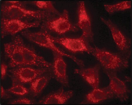
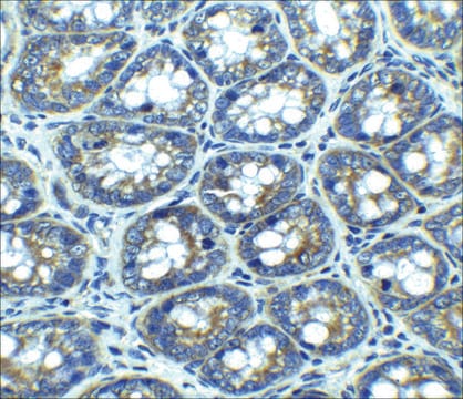
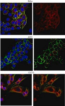
免疫细胞化学分析: 先前批次的抗体以 1:500 的稀释度在 NIH/3T3、A431 和 HeLa 细胞中检测到 LAMP-1。
该抗 LAMP-1(CD107a)抗体经过验证可用在 WB 和 IC 中检测 LAMP-1(CD107a)。

品質

通过蛋白质印迹在 EL4 细胞裂解液中进行了评估。

蛋白质印迹分析:1 µg/mL 的该抗体在 10 µg EL4 细胞裂解液中检测到 LAMP-1。

標靶描述

可观察到约95kDa的条带。 尽管计算得出的分子量为 45 kDa,但 LAMP-1 被高度糖基化,因此,预期典型的条带在 90-120 kDa 之间。在某些裂解液中可能会观察到未表征的条带。

外觀

亲和纯化
纯化的兔多克隆抗体,溶于含有0.1 M Tris-甘氨酸(pH 7.4)、150 mM NaCl和0.05%叠氮化钠的缓冲液中。

儲存和穩定性

自收到之日起,在2-8°C条件下可稳定保存1年。

分析報告

对照
EL4 细胞裂解液

其他說明

浓度:请参考批次特异性浓缩物的检验报告。

免責聲明

除非我们的产品目录或产品附带的其他公司文档另有说明,否则我们的产品仅供研究使用,不得用于任何其他目的,包括但不限于未经授权的商业用途、体外诊断用途、离体或体内治疗用途或任何类型的人类或动物食用或应用。

未找到適合的產品?  

試用我們的產品選擇工具.

儲存類別代碼

12 - Non Combustible Liquids

水污染物質分類(WGK)

WGK 1

閃點(°F)

Not applicable

閃點(°C)

Not applicable


分析證明 (COA)

輸入產品批次/批號來搜索 分析證明 (COA)。在產品’s標籤上找到批次和批號,寫有 ‘Lot’或‘Batch’.。

已經擁有該產品?

您可以在文件庫中找到最近購買的產品相關文件。

存取文件庫

Carolina Duarte et al.
Journal of cellular and molecular medicine, 26(10), 2841-2851 (2022-04-17)
Emerging studies indicate that intracellular eukaryotic ceramide species directly activate cathepsin B (CatB), a lysosomal-cysteine-protease, in the cytoplasm of osteoclast precursors (OCPs) leading to elevated RANKL-mediated osteoclastogenesis and inflammatory osteolysis. However, the possible impact of CatB on osteoclastogenesis elevated by
Tiago Ferreira et al.
Viruses, 12(10) (2020-10-25)
H-1 protoparvovirus (H-1PV) is a self-propagating virus that is non-pathogenic in humans and has oncolytic and oncosuppressive activities. H-1PV is the first member of the Parvoviridae family to undergo clinical testing as an anticancer agent. Results from clinical trials in
Yoav Charpak-Amikam et al.
Scientific reports, 7(1), 8661-8661 (2017-08-19)
Human cytomegalovirus (HCMV) is a major human pathogen, causing serious diseases in immunocompromised populations and congenially infected neonates. One of the main immune cells acting against the virus are Natural Killer (NK) cells. Killing by NK cells is mediated by
Lucas T Jae et al.
Science (New York, N.Y.), 344(6191), 1506-1510 (2014-06-28)
Lassa virus spreads from a rodent to humans and can lead to lethal hemorrhagic fever. Despite its broad tropism, chicken cells were reported 30 years ago to resist infection. We found that Lassa virus readily engaged its cell-surface receptor α-dystroglycan
Leonardo Ermini et al.
Frontiers in cell and developmental biology, 9, 652651-652651 (2021-05-22)
Aberrant ceramide build-up in preeclampsia, a serious disorder of pregnancy, causes exuberant autophagy-mediated trophoblast cell death. The significance of ceramide accumulation for lysosomal biogenesis in preeclampsia is unknown. Here we report that lysosome formation is markedly increased in trophoblast cells

文章

Autophagy is a highly regulated process that is involved in cell growth, development, and death. In autophagy cells destroy their own cytoplasmic components in a very systematic manner and recycle them.

我們的科學家團隊在所有研究領域都有豐富的經驗,包括生命科學、材料科學、化學合成、色譜、分析等.

聯絡技術服務