推薦產品
生物源
mouse
品質等級
抗體表格
purified antibody
抗體產品種類
primary antibodies
無性繁殖
LY1C6, monoclonal
形狀
liquid
包含
≤0.09% sodium azide as preservative
物種活性
rat
製造商/商標名
Calbiochem®
儲存條件
OK to freeze
avoid repeated freeze/thaw cycles
同型
IgG1
運輸包裝
wet ice
儲存溫度
−20°C
目標翻譯後修改
unmodified
基因資訊
rat ... Lamp1(25328)
一般說明
Protein G purified mouse monoclonal antibody. Recognizes the ~120 kDa Lamp1 protein.
Recognizes the ~120 kDa Lamp1 protein.
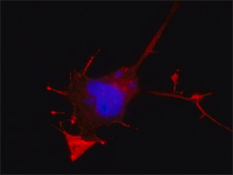
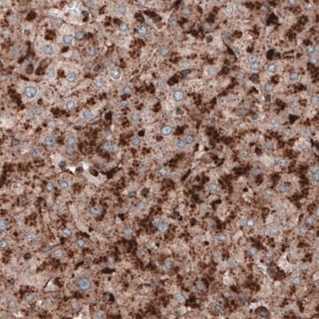
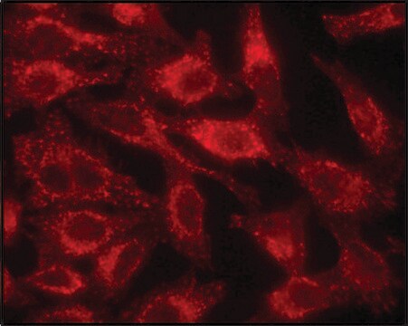
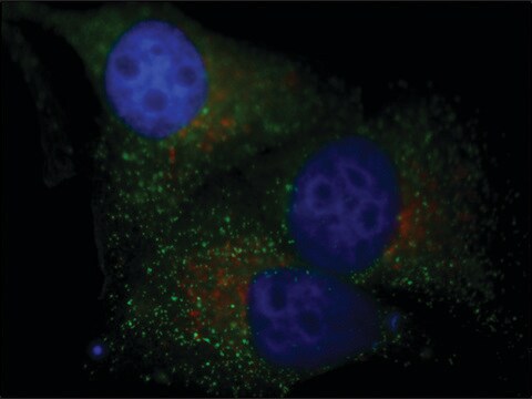
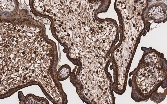
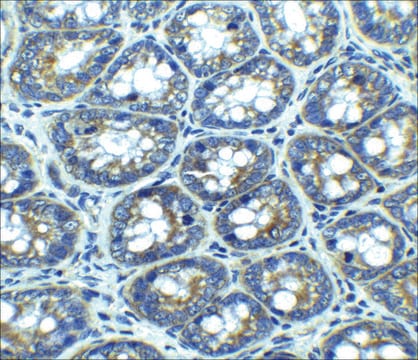
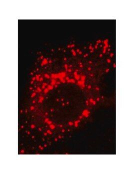
This Anti-Lamp1 Mouse mAb (LY1C6) is validated for use in Immunoblotting, Immunocytochemistry, Immunofluorescence, Immunoprecipitation for the detection of Lamp1.
免疫原
Rat
rat liver lysosomal membranes
應用
Immunoblotting (1 µg/ml, chemiluminescence)
Immunocytochemistry (1:100)
Immunofluorescence (see comments)
Immunoprecipitation (see comments)
Immunocytochemistry (1:100)
Immunofluorescence (see comments)
Immunoprecipitation (see comments)
包裝
Please refer to vial label for lot-specific concentration.
警告
Toxicity: Standard Handling (A)
外觀
In PBS containing 50% glycerol, pH 7.2.
重構
Following initial thaw, aliquot and freeze (-20°C).
分析報告
Positive Control
CHO-K1 cells
CHO-K1 cells
其他說明
Kannan, K. et al. 1996. Cell Immunol.171, 10.
Rohrer, J., et al. 1996. J. Cell Biol. 132, 565.
Howe et al. 1988. PNAS85, 7577.
Lewis et al. 1985. J. Cell Biol.100, 1839.
Rohrer, J., et al. 1996. J. Cell Biol. 132, 565.
Howe et al. 1988. PNAS85, 7577.
Lewis et al. 1985. J. Cell Biol.100, 1839.
This antibody has also been reported to work for immunofluorescence and immunoprecipitation. Antibody should be titrated for optimal results in individual systems.
法律資訊
CALBIOCHEM is a registered trademark of Merck KGaA, Darmstadt, Germany
未找到適合的產品?
試用我們的產品選擇工具.
儲存類別代碼
10 - Combustible liquids
水污染物質分類(WGK)
WGK 1
閃點(°F)
Not applicable
閃點(°C)
Not applicable
分析證明 (COA)
輸入產品批次/批號來搜索 分析證明 (COA)。在產品’s標籤上找到批次和批號,寫有 ‘Lot’或‘Batch’.。
Arvind Chhabra et al.
European journal of immunology, 34(10), 2824-2833 (2004-09-16)
Dendritic cells (DC) capture antigens from apoptotic and/or necrotic tumor cells and cross-present them to T cells, and various ways of delivering tumor antigens to DC in vitro and in vivo are being pursued. Since fusions of antigenic proteins with
Vanessa Ginet et al.
The American journal of pathology, 175(5), 1962-1974 (2009-10-10)
The multiplicity of cell death mechanisms induced by neonatal hypoxia-ischemia makes neuroprotective treatment against neonatal asphyxia more difficult to achieve. Whereas the roles of apoptosis and necrosis in such conditions have been studied intensively, the implication of autophagic cell death
Raymond E Hulse et al.
The Journal of neuroscience : the official journal of the Society for Neuroscience, 28(47), 12199-12211 (2008-11-21)
In brain, monomeric immunoglobin G (IgG) is regarded as quiescent and only poised to initiate potentially injurious inflammatory reactions via immune complex formation associated with phagocytosis and tumor necrosis factor alpha (TNF-alpha) production in response to disease. Using rat hippocampal
Julien Puyal et al.
Annals of neurology, 66(3), 378-389 (2009-06-25)
To evaluate the contributions of autophagic, necrotic, and apoptotic cell death mechanisms after neonatal cerebral ischemia and hence define the most appropriate neuroprotective approach for postischemic therapy. Rats were exposed to transient focal cerebral ischemia on postnatal day 12. Some
Vanessa Ginet et al.
Autophagy, 10(5), 846-860 (2014-03-29)
Neuronal autophagy is increased in numerous excitotoxic conditions including neonatal cerebral hypoxia-ischemia (HI). However, the role of this HI-induced autophagy remains unclear. To clarify this role we established an in vitro model of excitotoxicity combining kainate treatment (Ka, 30 µM)
文章
Autophagy is a highly regulated process that is involved in cell growth, development, and death. In autophagy cells destroy their own cytoplasmic components in a very systematic manner and recycle them.
我們的科學家團隊在所有研究領域都有豐富的經驗,包括生命科學、材料科學、化學合成、色譜、分析等.
聯絡技術服務

