推薦產品
品質等級
形狀
liquid
IVD
for in vitro diagnostic use
技術
microbe id | staining: suitable
pH值
3.7 (20 °C in H2O)
密度
1.01 g/cm3 at 20 °C
應用
clinical testing
diagnostic assay manufacturing
hematology
histology
儲存溫度
15-25°C
一般說明
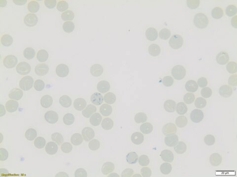
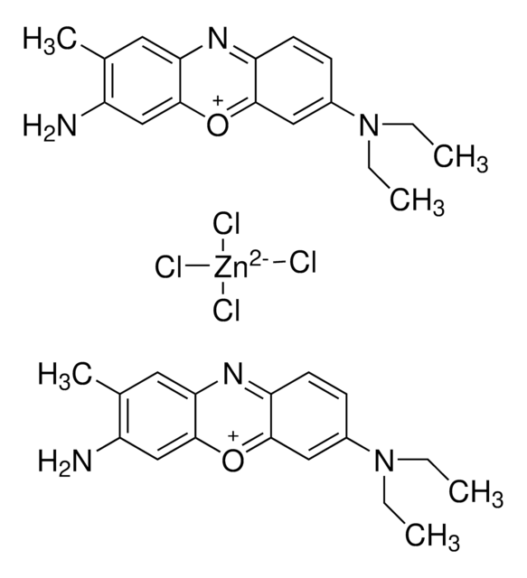
The Brilliant cresyl blue solution - for the staining of reticulocytes - for microscopy, is a ready-to-use solution used for human-medical cell diagnosis and serves the hematological investigation of sample material of human origin.
The dye stains the nucleic acids in the reticulocytes (Substantia granulofilamentosa), thus enabling reticulocytes to be morphologically distinguished from erythrocytes. The Substantia granulofilamentosa (ribonucleoproteins) can be rendered visible with fresh, non-fixed, young erythrocytes in a supervital staining procedure. Characterized by an intense black-blue hue, it is possible both to determine the number of the reticulocytes (visual microscopic counting) and also to visualize their morphological aspects. Four stages of substantia granulofilamentosa maturation can be distinguished depending on the stage of reticulocyte development: coiled skein (I), incomplete network (II), complete network (III) and granular form (IV). In peripheral blood the development stages III and IV are found most commonly.
The 100 ml bottle provides ca. 1000 applications. This product is registered as IVD and CE marked. For more details, please see instructions for use (IFU). The IFU can be downloaded from this webpage.
分析報告
Application test (Reticulocyte staining): passes test
儲存類別代碼
12 - Non Combustible Liquids
水污染物質分類(WGK)
WGK 2
閃點(°F)
Not applicable
閃點(°C)
Not applicable
分析證明 (COA)
輸入產品批次/批號來搜索 分析證明 (COA)。在產品’s標籤上找到批次和批號,寫有 ‘Lot’或‘Batch’.。
相關內容
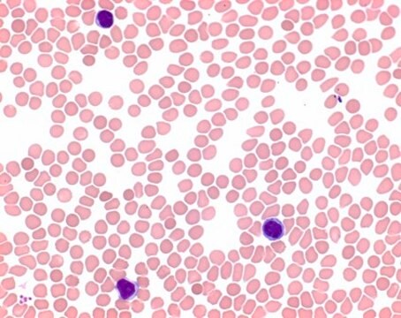
Learn about the clinical study of blood, blood-forming organs, and blood diseases including the treatment, prevention, and stains and dyes used in hematology testing.
Learn about the clinical study of blood, blood-forming organs, and blood diseases including the treatment, prevention, and stains and dyes used in hematology testing.
我們的科學家團隊在所有研究領域都有豐富的經驗,包括生命科學、材料科學、化學合成、色譜、分析等.
聯絡技術服務