MABN77
Anti-Potassium Channel Kv1.2 Antibody, clone K14/16
clone K14/16, from mouse
别名:
potassium voltage-gated channel, shaker-related subfamily, member 2, potassium voltage-gated channel subfamily A member 2, Voltage-gated potassium channel subunit Kv1.2, Voltage-gated potassium channel HBK5, potassium channel, Voltage-gated K(+) channel
登录查看公司和协议定价
所有图片(2)
About This Item
分類程式碼代碼:
12352203
eCl@ss:
32160702
NACRES:
NA.41
推荐产品
生物源
mouse
品質等級
抗體表格
purified antibody
抗體產品種類
primary antibodies
無性繁殖
K14/16, monoclonal
物種活性
rat
技術
immunofluorescence: suitable
immunohistochemistry: suitable
western blot: suitable
同型
IgG2bκ
NCBI登錄號
UniProt登錄號
運輸包裝
wet ice
目標翻譯後修改
unmodified
基因資訊
human ... KCNA2(3737)
一般說明
Potassium channel Kv1.2 is one of many different types of potassium channels in the cell. The Kv family of potassium channels seem to have a conserved homotetramer formation consisting of four voltage sensors and one pore domain. Kv1.2 cooperates with cortactin (an actin cytoskeleton-binding protein) and together they may have a part in the regulation of the ionic current.
免疫原
Recombinant protein corresponding to rat Kv1.2.
應用
Immunofluorescence Analysis: a previous lot of this antibody was used by an independent laboratory in IF. (Yang, J.W., et al. (2007). PNAS. 104(50):20055–20060.)
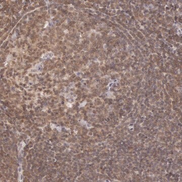
This Anti-Potassium Channel Kv1.2 Antibody, clone K14/16 is validated for use in IH, WB, IF for the detection of Potassium Channel Kv1.2.
品質
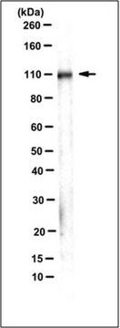
Evaluated by Western Blot in rat brain membrane tissue lysate.
Western Blot Analysis: 0.5 µg/mL of this antibody detected Kv1.2 on 10 µg of rat brain membrane tissue lysate.
Western Blot Analysis: 0.5 µg/mL of this antibody detected Kv1.2 on 10 µg of rat brain membrane tissue lysate.
標靶描述
~ 65 kDa observed
外觀
Format: Purified
其他說明
Concentration: Please refer to the Certificate of Analysis for the lot-specific concentration.
未找到合适的产品?
试试我们的产品选型工具.
儲存類別代碼
12 - Non Combustible Liquids
水污染物質分類(WGK)
WGK 1
閃點(°F)
Not applicable
閃點(°C)
Not applicable
De-En Xu et al.
Cell adhesion & migration, 8(4), 396-403 (2014-12-09)
Amyloid precursor protein (APP), commonly associated with Alzheimer disease, is upregulated and distributes evenly along the injured axons, and therefore, also known as a marker of demyelinating axonal injury and axonal degeneration. However, the physiological distribution and function of APP
Nikki A McLean et al.
PloS one, 9(10), e110174-e110174 (2014-10-14)
Rapid and efficient axon remyelination aids in restoring strong electrochemical communication with end organs and in preventing axonal degeneration often observed in demyelinating neuropathies. The signals from axons that can trigger more effective remyelination in vivo are still being elucidated.
我们的科学家团队拥有各种研究领域经验,包括生命科学、材料科学、化学合成、色谱、分析及许多其他领域.
联系技术服务部门


