Skip to Content
Merck
All Photos(1)

Key Documents

F6182

Sigma-Aldrich

Fluoroshield

liquid

Sign Into View Organizational & Contract Pricing


About This Item

UNSPSC Code:
12171500
NACRES:
NA.47

Product Name

Fluoroshield, histology mounting medium

Quality Level

form

liquid

technique(s)

immunofluorescence: suitable

pH range

7.8-8.2

solubility

water: soluble

application(s)

diagnostic assay manufacturing
hematology
histology

storage temp.

2-8°C

General description

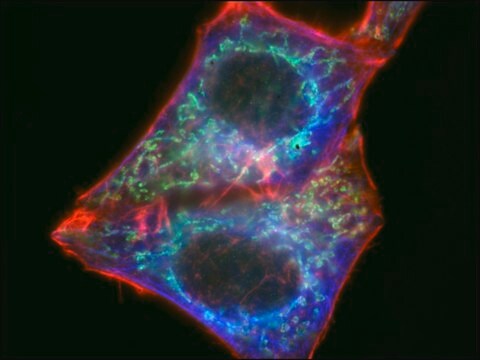
Fluoroshield is an aqueous mounting medium for preserving fluorescence of tissue and cell smears. This unique formula not only enables prolonged fluorescence, but also prevents rapid photobleaching (fading) of fluorochromes including FITC, Texas Red®, AMCA, Cy®, Cy3, Cy5, Alexa Fluor 488, Alexa Fluor 594, Green fluorescent protein (GFP), tetramethylrhodamine, R-phycoerythrin (R-PE), phycocyanin (PC), and allophycocyanin (APC).

Application

Fluoroshield has been used:
  • as a mounting medium for cultured megakaryocytes and immunostained platelets for confocal microscopy studies
  • as a mounting medium for 4′,6-diamidino-2-phenylindole (DAPI) stained heart sections for immunofluorescence studies
  • to preserve fluorescence of C6 cell smears

Linkage

Fluoroshield is an aqueous-based mounting medium comparable to Gel Mount.

Legal Information

ALEXA FLUOR is a trademark of Life Technologies
Cy is a registered trademark of Cytiva
Cy3 is a trademark of Cytiva
FluoroShield is a trademark of ImmunoBioScience Corp.
Gel Mount is a trademark of Biomeda Corp.
Texas Red is a registered trademark of Life Technologies

related product

Product No.
Description
Pricing

Storage Class Code

10 - Combustible liquids

WGK

WGK 1

Flash Point(F)

Not applicable

Flash Point(C)

Not applicable


Choose from one of the most recent versions:

Certificates of Analysis (COA)

Lot/Batch Number

Don't see the Right Version?

If you require a particular version, you can look up a specific certificate by the Lot or Batch number.

Already Own This Product?

Find documentation for the products that you have recently purchased in the Document Library.

Visit the Document Library

Chenchen Yang et al.
Scientific reports, 8(1), 16359-16359 (2018-11-07)
Visibly transparent luminescent solar concentrators (TLSC) have the potential to turn existing infrastructures into net-zero-energy buildings. However, the reabsorption loss currently limits the device performance and scalability. This loss is typically defined by the Stokes shift between the absorption and
Effects of dexamethasone on C6 cell proliferation, migration and invasion through the upregulation of AQP1
Guan Y, et al.
Oncology Letters, 15(5), 7595-7602 (2018)
F Lodola et al.
Science advances, 5(9), eaav4620-eaav4620 (2019-10-11)
The control of stem and progenitor cell fate is emerging as a compelling urgency for regenerative medicine. Here, we propose a innovative strategy to gain optical control of endothelial colony-forming cell fate, which represents the only known truly endothelial precursor
Douglas H Campbell et al.
PloS one, 13(4), e0196017-e0196017 (2018-04-20)
While measurement of serum prostate specific antigen (PSA) is an important screening tool for prostate cancer, new biomarkers are necessary for better discrimination between presence and absence of disease. The MIL-38 monoclonal antibody is specific for the membrane glycoprotein glypican
Venugopal Thayanithy et al.
Cell communication and signaling : CCS, 15(1), 46-46 (2017-11-15)
Tunneling nanotubes (TNTs) are naturally-occurring filamentous actin-based membranous extensions that form across a wide spectrum of mammalian cell types to facilitate long-range intercellular communication. Valid assays are needed to accurately assess the downstream effects of TNT-mediated transfer of cellular signals

Articles

The Human Protein Atlas Program has carefully selected three different human cell lines, A-431 epidermoid carcinoma, U-251 MG glioblastoma and U-205 osteosarcoma, for organelle mapping of the proteome. As Prestige Antibodies are studied by immunofluorescence (IF) staining, three well-characterized organelle markers for nuclei, microtubules and endoplasmic reticulum are used correspondingly as specific probes. The high-resolution confocal images are annotated based on sub-cellular localization. In addition, staining intensity and characteristics are also part of an overall assessment as well as comparison to literature (if available).

Our team of scientists has experience in all areas of research including Life Science, Material Science, Chemical Synthesis, Chromatography, Analytical and many others.

Contact Technical Service