All Photos(1)
About This Item
UNSPSC Code:
41106514
biological source:
mouse brain
growth mode:
Adherent
karyotype:
Not specified
morphology:
Neuronal
products:
Not specified
receptors:
Not specified
Recommended Products
Product Name
C17.2, 07062902
biological source
mouse brain
growth mode
Adherent
karyotype
Not specified
morphology
Neuronal
products
Not specified
receptors
Not specified
technique(s)
cell culture | mammalian: suitable
shipped in
dry ice
storage temp.
−196°C
Cell Line Origin
Mouse multipotent neural progenitor or stem-like cells
Cell Line Description
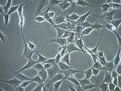
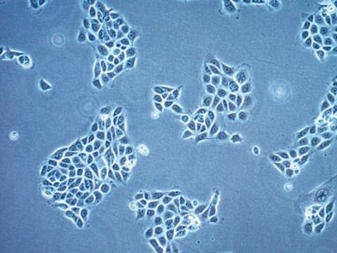
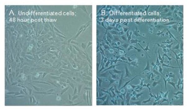
An immortalised mouse neural progenitor cell line capable of differentiation in vitro. The cell line was established by retorviral-mediated transduction of the avian myc oncogene into mitotic progenitor cells of neonatal mouse cerrebellum. Mouse strain CD1 x C57BL/6. This cell line is a valuable tool for in vitro and in vivo studies aimed at understanding the control of cell fate and differentiation of neural progenitors. The MMLV retrovirus vector used for the immortalisation process contained a neo resistance gene transcribed from an internal SV40 promoter. Therefore the cells are neo resistant. The morphology of the cells may change over time. Cells plated at low density may tend to become more process bearing whereas those plated more densely may tend to become flat and non-process bearing.
Culture Medium
DMEM + 2 mM Glutamine + 10% Fetal Bovine Serum (FBS). Flasks should be pre-coated with poly-L-lysine at 10 μg/ml in sterile distilled water.
Subculture Routine
Feed cells weekly with 50% conditioned medium and 50% fresh medium or split 1:10-1:20 in fresh medium using trypsin/EDTA. 5% CO2; 37 °C. Split sub-confluent cultures 1:10 - 1:20, i.e., seeding at 2-4 x 10,000 cells/cm2. Flasks should be pre-coated with poly-L-lysine at 10 μg/ml in sterile distilled water. Cells can be split as dilute as 1:50, but the phenotype may change, i.e., cells may appear flat with an epithelial-like morphology which usually do not stain for neurofilament or GFAP.
Other Notes
Additional freight & handling charges may be applicable for Asia-Pacific shipments. Please check with your local Customer Service representative for more information.
Choose from one of the most recent versions:
Certificates of Analysis (COA)
Lot/Batch Number
Sorry, we don't have COAs for this product available online at this time.
If you need assistance, please contact Customer Support.
Already Own This Product?
Find documentation for the products that you have recently purchased in the Document Library.
Our team of scientists has experience in all areas of research including Life Science, Material Science, Chemical Synthesis, Chromatography, Analytical and many others.
Contact Technical Service


