COLLA-RO
Roche
Collagenase A
from Clostridium histolyticum
About This Item
Recommended Products
biological source
Clostridium histolyticum
Quality Level
sterility
non-sterile
form
lyophilized
specific activity
>0.15 U/mg lyophilizate (Collagenase activity)
packaging
pkg of 100 mg (10103578001)
pkg of 2.5 g (11088793001)
pkg of 500 mg (10103586001)
manufacturer/tradename
Roche
concentration
1 mg/mL (0.1%, w/v)
color
brown
optimum pH
6.0-8.0
solubility
water: soluble
suitability
suitable for PCR
NCBI accession no.
UniProt accession no.
application(s)
cell analysis
life science and biopharma
sample preparation
foreign activity
Clostripain 9.489 U/mg
Proteases 6.067 U/mg (Azocoll )
Trypsin 0.144 U/mg (with BAEE)
storage temp.
2-8°C
Gene Information
Clostridium histolyticum ... COLA_1(1668506084)
General description
Specificity
Enzyme activity:
Collagenase activity: >0.15U/mg (according to Wünsch) (+25°C, 4-phenyl-azobenzyl-oxycarbonyl-Pro-Leu-Gly-Pro-D-Arg as the substrate)
Contaminating enzyme activities: trypsin, clostripain, and neutral proteolytic activity
Collagenase A has a balanced ratio of enzyme activities.
Application
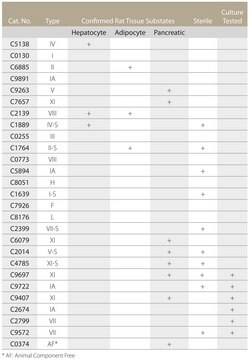
- Disaggregation of many types of tissues (e.g., lung, heart, muscle, bone, adipose tissue, liver, kidney, cartilage, mammary gland, placentae, blood vessels, brain, tumors, umbilical cord)
- preparation of single-cell suspensions for the establishment of primary cell culture systems. Collagenase A is recommended when yield, viability, and functionality are important.
- preparation of cells from many types of tissue, such as hepatocytes, adipocytes, pancreatic islets, epithelial cells, muscle cells, endothelial cells, etc. However, the suitability of each lot of the enzyme for disruption of a particular tissue should be determined empirically.
Unit Definition
Lyophilizate, nonsterile
Frequently, collagenase activities are given in Mandl units (1 μmol leucine liberated from collagen in 5 hours at +37 °C).
Unfortunately, there is no consistent conversion factor between the two units of activity, since the Mandl unit depends, in part, on the concentration of contaminating proteases in the collagenase preparation, an indefinable variable. A purer collagenase preparation would actually give a lower specific activity in Mandl units than a crude preparation. Clostridium preparations typically give conversion factors of approximately 1:1800 (e.g., a particular lot of Clostridium collagenase contained approximately 0.15 Wünsch U/mg and 250 Mandl U/mg).
Preparation Note
Inhibitors:
Collagenase inhibitors: EDTA, EGTA, Cys, His, DTT, 2-mercapto-ethanol
Collagenase is not inhibited by serum.
Clostripain inhibitors: TLCK
Trypsin inhibitors: aprotinin, trypsin inhibitor (egg white, soybean), serum
Working concentration: 0.5 to 2.5 mg/ml
Storage conditions (working solution): -15 to -25 °C
Roche recommends reconstituting only the amount of lyophilizate needed for immediate use. The reconstituted solution can be stored at -15 to -25 °C for up to one week. Avoid repeated freezing and thawing since activity decreases after reconstitution.
Reconstitution
Storage and Stability
Other Notes
Signal Word
Danger
Hazard Statements
Precautionary Statements
Hazard Classifications
Eye Irrit. 2 - Resp. Sens. 1 - Skin Irrit. 2 - STOT SE 3
Target Organs
Respiratory system
Storage Class Code
11 - Combustible Solids
WGK
WGK 1
Flash Point(F)
does not flash
Flash Point(C)
does not flash
Certificates of Analysis (COA)
Search for Certificates of Analysis (COA) by entering the products Lot/Batch Number. Lot and Batch Numbers can be found on a product’s label following the words ‘Lot’ or ‘Batch’.
Already Own This Product?
Find documentation for the products that you have recently purchased in the Document Library.
Customers Also Viewed
Articles
Enzyme Explorer Key Resource: Collagenase Guide.Collagenases, enzymes that break down the native collagen that holds animal tissues together, are made by a variety of microorganisms and by many different animal cells.
Our team of scientists has experience in all areas of research including Life Science, Material Science, Chemical Synthesis, Chromatography, Analytical and many others.
Contact Technical Service


