Skip to Content
Merck
All Photos(2)

Documents

MAB3822-C

Sigma-Aldrich

Anti-Ezrin Antibody, clone 4A5, Ascites Free

clone 4A5, from mouse

Synonym(s):

Cytovillin, Villin-2, p81, Ezrin

Sign Into View Organizational & Contract Pricing


About This Item

UNSPSC Code:
12352203
eCl@ss:
32160702
NACRES:
NA.41

biological source

mouse

Quality Level

antibody form

purified immunoglobulin

antibody product type

primary antibodies

clone

4A5, monoclonal

species reactivity

human, rat, rabbit, mouse

technique(s)

electron microscopy: suitable
immunocytochemistry: suitable
immunohistochemistry: suitable
immunoprecipitation (IP): suitable
western blot: suitable

isotype

IgG1κ

NCBI accession no.

UniProt accession no.

shipped in

wet ice

target post-translational modification

unmodified

Gene Information

human ... EZR(7430)

General description

Ezrin (UniProt Q8HZQ5; also known as Cytovillin, Villin-2, p81) is encoded by the EZR (also known as VIL2) gene (Gene ID 100008846) in Oryctolagus cuniculus (Rabbit) species. Ezrin, radixin, and moesin are the three ERM protein family members that crosslink actin filaments with plasma membranes and play an important role in the cell cortex architecture. In its inactive state, ezrin is located in the cytosol in a conformation involving intramolecular interaction between its N- and C-terminal. Ezrin is activated by phosphorylation, which causes a conformation change that disrupts the head-to-tail intramolecular interaction. Upon activation, ezrin binds to the cytoplasmic tails of multiple adhesion and signaling membrane molecules, such as CD44, growth factor receptors, and vascular adhesion molecule (VCAM1). The initiator methionine of ezrin is removed posttranslationally to yield the mature protein (a.a. 2-586) with an N-termina FERM domain (a.a. 2-295) that mediates interaction with microtubules.

Specificity

Clone 4A5 reacted with both dentured and non-denatured Ezrin (Hanzel, D.K., et al. (1989). Am. J. Physiol. 256(6 Pt 1):G1082-G1089).

Immunogen

Purified Ezrin from rabbit gastric parietal cells.

Application

Anti-Ezrin Antibody, clone 4A5, Ascites Free is an antibody against Ezrin for use in Western Blotting, Immunohistochemistry, Immunoprecipitation, Electron Microscopy, Immunocytochemistry.
Research Category
Cell Structure
Research Sub Category
Adhesion (CAMs)
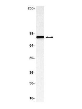
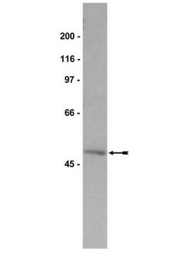
Western Blotting Analysis: 0.5 µg/mL of this antibody from a representative lot detected Ezrin in 10 µg of HeLa cell lysate.
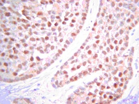
Immunohistochemistry Analysis: A representative lot detected a higher Ezin immunoreactivity in paraffin-embedded human medulloblastoma sections when compared with normal cerebellum sections (Haeberle, H., et al. (2012). Neoplasia. 14(7):666-669).
Immunohistochemistry Analysis: A representative lot immunostained parietal cells in rabbit gastric mucosa (Hanzel, D.K., et al. (1989). Am. J. Physiol. 256(6 Pt 1):G1082-G1089).
Immunoprecipitation Analysis: A representative lot immunoprecipitated Ezin from human A431 cells. The immunoprecipitated Ezrin was phosphorylated on Ser66 from dbcAMP‐treated, but not cAMP‐treated, A431 cells (Wang, S., et al. (2009). Cell Res. 19(12):1350-1362).
Electron Microscopy Analysis: A representative lot detected Ezrin on microvilli of the intracellular canaliculi (IC) and the subapical region of resting parietal cells in Lowicryl HM20-embedded rabbit gastric gland ultrathin sections (Sawaguchi, A., et al. (2004). J. Histochem. Cytochem. 52(1): 77–86).
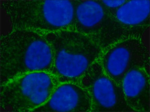
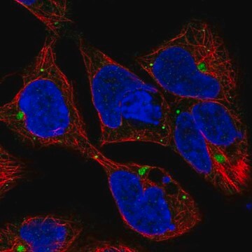
Immunocytochemistry Analysis: A representative lot detected exogenously expressed human GST fusion protein by fluorescent immunocytochemistry staining of formaldehyde-fixed, Triton X-100-permeabilized rabbit gastric parietal cells (Zhou, R., et al. (2003). J. Biol. Chem. 278(37):35651-35659).
Western Blotting Analysis: A representative lot detected both the endogenous rabbit Ezrin as well as exogenously expressed human Ezrin GST fusion proteins in transfected rabbit gastric parietal cells (Zhou, R., et al. (2003). J. Biol. Chem. 278(37):35651-35659).
ELISA Analysis: Clone 4A5 hybridoma culture supernatant was screened for its immunoreactivity by ELISA and found to react with both dentured and non-denatured 80 kDa Ezrin preparations from rabbit gastric glands/mucosa vesicular fractions (Hanzel, D.K., et al. (1989). Am. J. Physiol. 256(6 Pt 1):G1082-G1089).

Quality

Evaluated by Western Blotting in L6 rat myoblast lysate.

Western Blotting Analysis: 0.5 µg/mL of this antibody detected Ezrin in 10 µg of L6 rat myoblast lysate.

Target description

~75 kDa observed. 69.41 kDa (Human/UniProt P15311 & Mouse/P26040), 69.39 kDa (Rat/UniProt P31977), and 69.22 kDa (Rabbit/Q8HZQ5) calculated. Uncharacterized band(s) may appear in some lysates.

Physical form

Format: Purified
Protein G Purified
Purified mouse monoclonal IgG1κ antibody in buffer containing 0.1 M Tris-Glycine (pH 7.4), 150 mM NaCl with 0.05% sodium azide.

Storage and Stability

Stable for 1 year at 2-8°C from date of receipt.

Other Notes

Concentration: Please refer to lot specific datasheet.

Disclaimer

Unless otherwise stated in our catalog or other company documentation accompanying the product(s), our products are intended for research use only and are not to be used for any other purpose, which includes but is not limited to, unauthorized commercial uses, in vitro diagnostic uses, ex vivo or in vivo therapeutic uses or any type of consumption or application to humans or animals.

Not finding the right product?  

Try our Product Selector Tool.

Storage Class Code

12 - Non Combustible Liquids

WGK

WGK 1

Flash Point(F)

Not applicable

Flash Point(C)

Not applicable


Certificates of Analysis (COA)

Search for Certificates of Analysis (COA) by entering the products Lot/Batch Number. Lot and Batch Numbers can be found on a product’s label following the words ‘Lot’ or ‘Batch’.

Already Own This Product?

Find documentation for the products that you have recently purchased in the Document Library.

Visit the Document Library

M Motiur Rahman et al.
Cells, 10(3) (2021-04-04)
In Inflammatory Bowel Disease (IBD), malabsorption of electrolytes (NaCl) results in diarrhea. Inhibition of coupled NaCl absorption, mediated by the dual operation of Na:H and Cl:HCO3 exchangers on the brush border membrane (BBM) of the intestinal villus cells has been
M Motiur Rahman et al.
International journal of molecular sciences, 22(8) (2021-05-01)
Electrolytes (NaCl) and fluid malabsorption cause diarrhea in inflammatory bowel disease (IBD). Coupled NaCl absorption, mediated by Na+/H+ and Cl-/HCO3- exchanges on the intestinal villus cells brush border membrane (BBM), is inhibited in IBD. Arachidonic acid metabolites (AAMs) formed via
Molly Butts et al.
The Journal of nutrition, 150(4), 747-755 (2019-11-27)
Chronic alcohol use often leads to malnutrition. However, how the intestinal absorption of nutrients such as glucose may be affected during moderate ethanol use has not been investigated. Glucose is absorbed via sodium (Na)-dependent glucose co-transport (SGLT1; SLC5A1) along the

Our team of scientists has experience in all areas of research including Life Science, Material Science, Chemical Synthesis, Chromatography, Analytical and many others.

Contact Technical Service